An echocardiogram — often called an "echo" — is a non-invasive, radiation-free ultrasound scan of the heart. It shows the heart's chambers, valves, and blood flow in real time, and is the most widely used imaging test in cardiology. An echo can diagnose a new heart problem, track how a known condition is progressing, or confirm that the heart is structurally normal.
What Is an Echocardiogram?
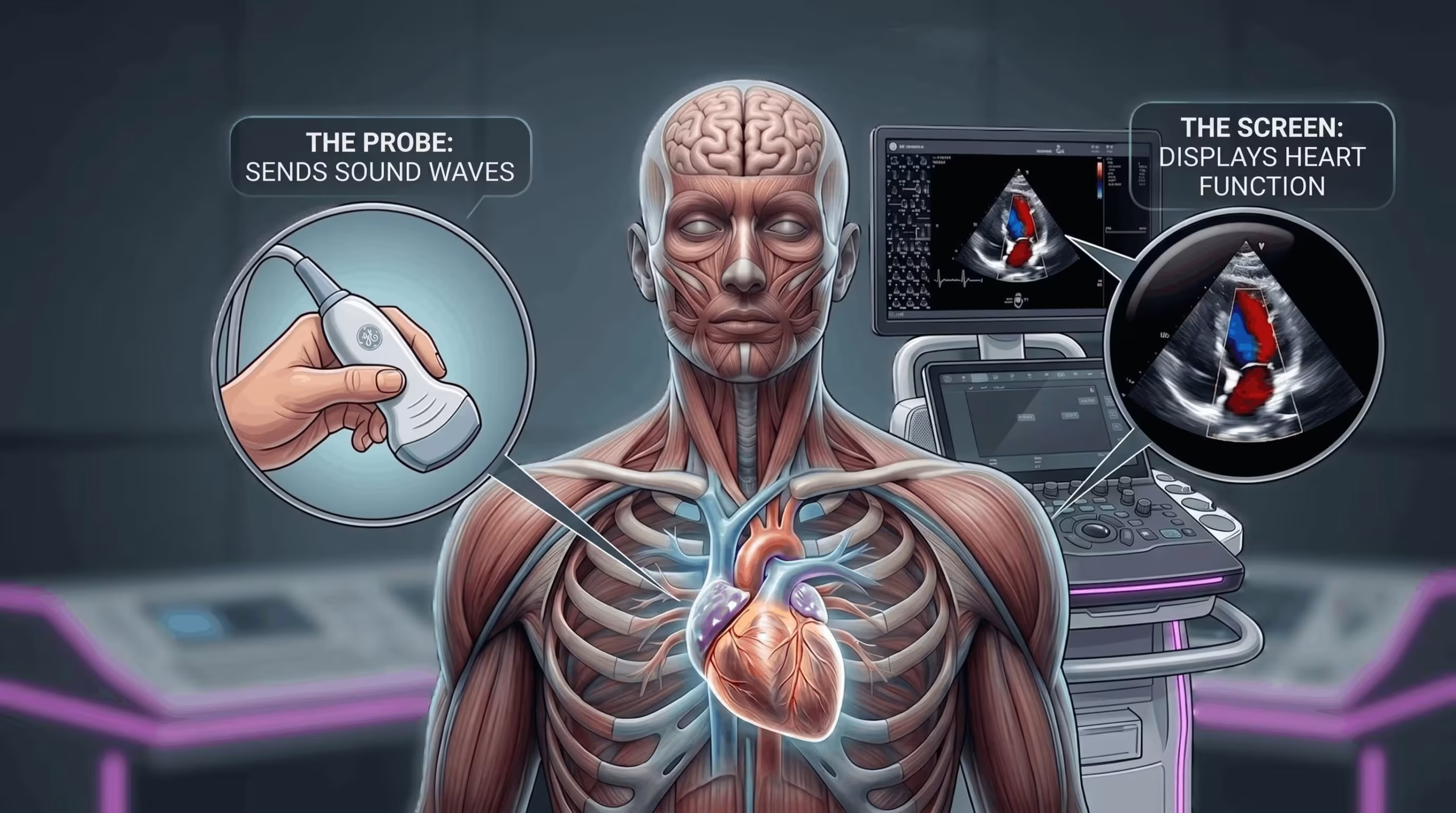
An echocardiogram uses ultrasound — high-frequency sound waves — to produce moving images of the heart. A sonographer places a handheld probe (transducer) on the chest, and the reflected sound waves are processed into real-time images of the heart's chambers, valves, blood flow, and great vessels.
The scan involves no radiation, no injections in most cases, and no preparation. It is performed in an outpatient setting and usually takes 30–45 minutes. Its safety, accessibility, and diagnostic power have made echocardiography the most widely used cardiac imaging test in the world. For clinicians wanting technical detail on measurements and reporting, see our echocardiography reference hub.
What Does an Echocardiogram Show?
An echocardiogram provides a real-time window into the structure and function of the heart. The main findings it can identify are:
- Chamber size and shape — whether the heart is enlarged, thickened, or has abnormal geometry
- Pumping function — how strongly the left and right ventricles contract, commonly reported as ejection fraction
- Valve structure and flow — whether valves open and close normally, and whether blood is leaking (regurgitation) or obstructed (stenosis)
- Fluid around the heart — pericardial effusion, and whether it is causing haemodynamic compromise
- Pressure clues — indirect estimates of pulmonary artery pressures and filling pressures from Doppler measurements
- Clots and masses — intracardiac thrombus, infective vegetations, or tumours
An echocardiogram cannot directly visualise the coronary arteries or confirm an acute heart attack — other tests are used for those questions.
Echocardiogram vs ECG: What's the Difference?
These two tests sound similar but answer completely different questions. An ECG (electrocardiogram) records the heart's electrical activity through small stickers on the chest — it diagnoses rhythm and conduction disorders but tells you nothing about structure. An echocardiogram uses ultrasound to show the heart's physical structure and function, but does not measure electrical activity. The two are complementary and are often ordered together.
| Feature | ECG | Echocardiogram |
|---|---|---|
| What it measures | Electrical activity | Structure and function |
| Technology | Surface electrodes | Ultrasound imaging |
| Diagnoses | Arrhythmias, conduction disease, some ischaemia | Valve disease, cardiomyopathy, effusion, LV function, masses |
| Duration | 1–2 minutes | 30–45 minutes |
| Radiation | None | None |
Types of Echocardiogram
Not every echo is the same. The choice depends on the clinical question, body habitus, and which structures need to be imaged.
| Type | How it's done | Best for |
|---|---|---|
| Transthoracic echo (TTE) | Probe on the chest wall | First-line for almost all clinical questions |
| Transoesophageal echo (TOE) | Probe passed into the oesophagus under light sedation | Left atrial appendage, prosthetic valves, endocarditis, cardiac masses |
| Stress echocardiogram | TTE performed with exercise or pharmacological stress | Inducible ischaemia, exertional symptoms |
| Contrast echocardiography | Intravenous ultrasound contrast agent | Poor image quality, suspected LV thrombus |
| 3D echocardiography | Real-time 3D reconstruction | Detailed valve anatomy, pre-operative planning |
At Heartcare Sydney we offer transthoracic echocardiography and stress echocardiography on site. TOE and specialised 3D imaging are arranged through our affiliated hospitals when clinically indicated.
What to Expect During an Echocardiogram
The test is painless, non-invasive, and typically takes less than an hour. Here is what happens, step by step.
Before the test
You remove clothing from the upper body and lie on an examination table. Small ECG stickers are placed on the chest to monitor your rhythm during the scan.
Gel is applied
A water-based ultrasound gel is applied to the chest to help the probe glide smoothly and improve image clarity. It may feel mildly cold, but is harmless.
Image acquisition
The sonographer glides the probe across several positions on the chest, occasionally asking you to shift position or hold your breath briefly for clearer views.
Duration
Most transthoracic scans take 30–45 minutes. Stress and transoesophageal echoes take longer because of preparation and recovery time.
After the test
The gel is wiped off and you return to normal activity immediately. Images are reviewed by the cardiologist, who discusses the results with you or your referring doctor.
When Is an Echocardiogram Recommended?
An echocardiogram is recommended whenever structural or functional information about the heart is needed. The most common reasons fall into four groups: new symptoms, known structural heart disease, acute or infective conditions, and monitoring across chronic or specialised care.
Symptoms
Heart murmur
An unusual sound detected on examination. Echo identifies its cause — most often a valve abnormality — or confirms the murmur is innocent.
Chest pain or breathlessness
To assess whether heart failure, valve disease, or other structural cause is contributing to the symptoms.
Palpitations or irregular rhythm
Echo assesses the underlying structural substrate — chamber size, LV function, valve disease — which influences rhythm management.
Structural heart conditions
Valvular heart disease
Stenosis, regurgitation, or prolapse of any of the four heart valves. Echo is the gold standard for assessing severity.
Cardiomyopathy
Diseases of the heart muscle affecting its pumping ability — dilated, hypertrophic, or restrictive forms.
Congenital heart disease
Defects present from birth, identified in children or in adults. Echo diagnoses, monitors, and guides intervention.
Acute or infective
Pericardial effusion or tamponade
Fluid collecting around the heart. Echo identifies the effusion and assesses whether it is causing haemodynamic compromise.
Infective endocarditis
Infection of a valve or intracardiac device, confirmed by vegetations on echocardiography, often requiring TOE.
Pulmonary hypertension
Elevated pressure in the lung circulation. Echo is the first-line non-invasive screening tool.
Monitoring and specialised
Monitoring known heart disease
Tracking valve disease, LV function, or cardiomyopathy over time. Serial scans guide timing of intervention.
Before non-cardiac surgery
In higher-risk patients, to assess baseline cardiac function and exclude significant valvular or systolic dysfunction.
Cancer therapy surveillance
Detecting cardiotoxicity from chemotherapy or targeted agents. See our dedicated page on echocardiography in cardio-oncology.
Special Situations Where Echo Plays a Central Role
Three areas where echocardiography is central enough to deserve its own article. Each is covered in depth on a dedicated page.
Echocardiography in stroke evaluation
Around 1 in 4 ischaemic strokes originates from the heart. Echo identifies clots, valve disease, PFO, and other cardiac sources that change treatment.
Echocardiography in cardio-oncology
Several cancer therapies can damage the heart. Echo is the primary tool for baseline risk, early detection of cardiotoxicity, and long-term surveillance.
Cardiac masses on echocardiography
Rare but important. Two clinical cases — a benign atrial myxoma and metastatic melanoma — illustrate the role of echo in detection and characterisation.
- An echocardiogram is a non-invasive ultrasound scan of the heart — no radiation, no preparation, no recovery.
- It shows chamber size, pumping function, valve behaviour, pericardial fluid, pressure clues, and intracardiac masses.
- TTE is the first-line test; TOE, stress echo, contrast, and 3D echo extend the toolkit for specific clinical questions.
- Echo and ECG are complementary — one shows structure, the other shows electrical activity.
- Indications fall into four broad groups: symptoms, structural disease, acute or infective conditions, and monitoring contexts.
- Most scans take 30–45 minutes and involve no recovery period.
Frequently Asked Questions
What does an echocardiogram show?
The size and shape of the heart chambers, how strongly the heart is pumping, the function of the valves, the presence of fluid around the heart, indirect estimates of pressures, and abnormal structures such as clots or masses.
Is an echocardiogram painful?
No. The probe is pressed firmly on the chest, which may feel like mild pressure, but there is no discomfort of note and no needles for a standard scan.
How long does it take?
Most transthoracic scans take 30–45 minutes. Stress and transoesophageal echoes take longer because of preparation and recovery.
Do I need to prepare for the scan?
Not for a standard TTE. For a TOE, you'll be asked not to eat or drink for several hours beforehand because light sedation is used. For a stress echo, wear clothing suitable for exercise.
Can an echocardiogram detect a heart attack?
It cannot detect a heart attack directly, but it can show the consequences — a wall that isn't contracting normally, a clot in the left ventricle, or reduced overall pumping function. Diagnosis of an acute heart attack relies on ECG and blood tests.
Is an echocardiogram bulk billed in Australia?
At Heartcare Sydney, echocardiograms can be bulk billed for eligible patients with a valid referral. Stress echoes are not bulk billed. Consultations are privately billed with a Medicare rebate. A referral is required for all cardiology services.
How is an echocardiogram different from an ECG?
An ECG records the heart's electrical activity through stickers on the skin and diagnoses rhythm problems. An echocardiogram uses ultrasound to show structure and function. They answer different questions and are often ordered together.
Conclusion
Echocardiography is a cornerstone of modern cardiology — safe, widely available, radiation-free, and remarkably informative. It underpins the diagnosis of most structural heart disease and the monitoring of heart function during and after treatment. For most questions about the heart, an echocardiogram is the first test ordered, and often the most informative.
For more specialised applications of echocardiography, see our dedicated pages on stroke evaluation, cardio-oncology, and cardiac masses.