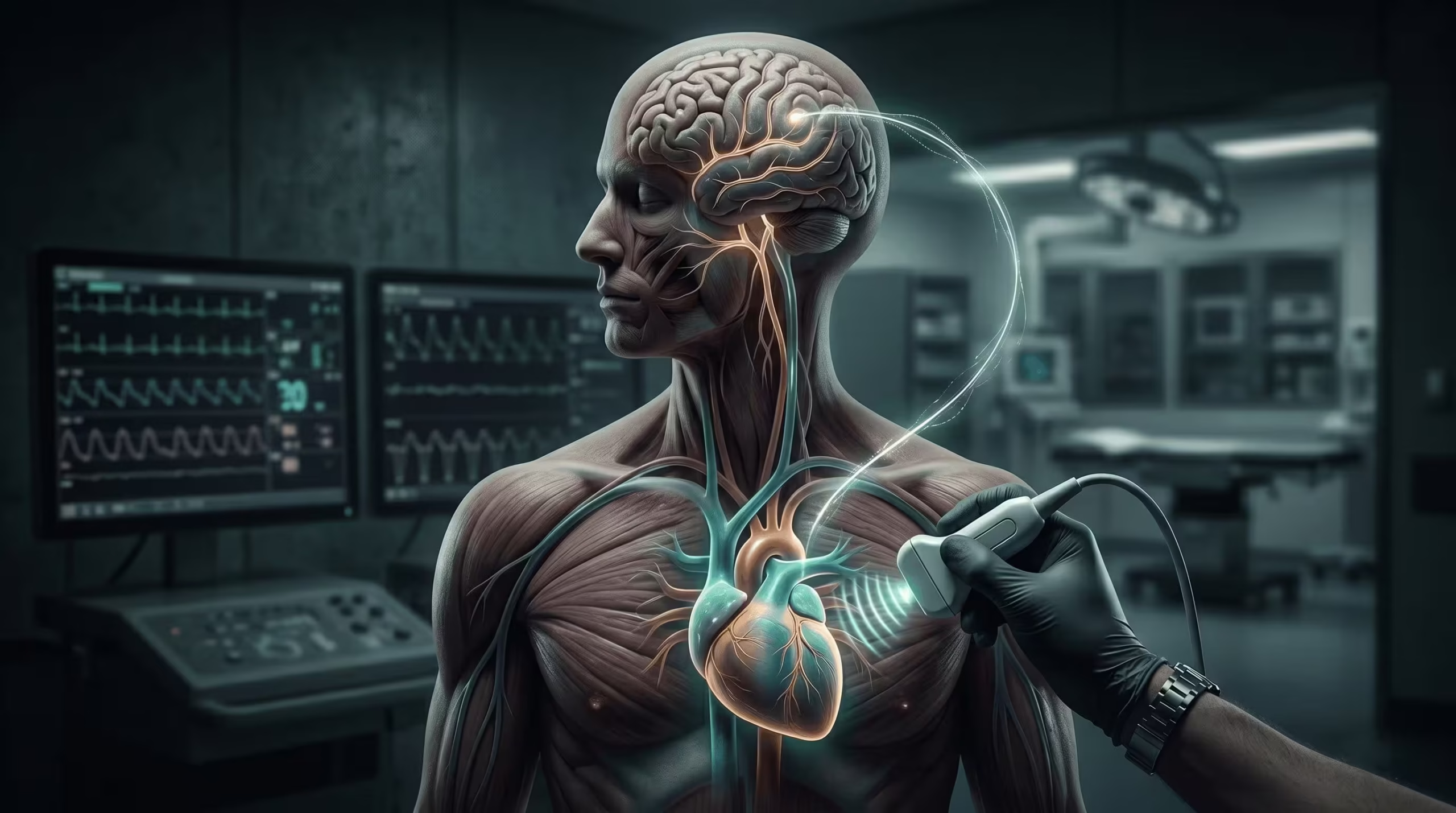
Approximately 20–25% of ischaemic strokes are cardioembolic — caused by clots, masses, or structural abnormalities that originate in the heart and travel to the brain. Identifying a cardiac source matters because it changes treatment: most cardioembolic strokes require anticoagulation rather than antiplatelet therapy, and some findings trigger specific interventions such as PFO closure, valve surgery, or tumour resection. Echocardiography is the cornerstone investigation for this workup.
Why an Echocardiogram Matters After a Stroke
An echocardiogram helps answer three questions that directly change your treatment:
- Did a clot come from the heart?
- Is there a structural problem that increases future stroke risk?
- Would the finding change your treatment — for example, anticoagulation rather than aspirin?
When Is an Echo Indicated After Stroke?
Not every ischaemic stroke requires an echocardiogram. Current AHA/ASA guidance recommends echo when a cardioembolic source is clinically suspected and when the result would change management. Certain clinical features raise that suspicion substantially.
Features that suggest a cardioembolic source
- Sudden onset with maximal deficit at presentation
- Cortical (not lacunar) infarct pattern
- Multiple or bilateral infarcts, or simultaneous cortical and cerebellar involvement
- No significant large-vessel disease on CTA or MRA
- Known cardiac risk factors: AF, recent MI, reduced ejection fraction, prosthetic valve, endocarditis
Timing is usually within days to weeks of presentation once the patient is haemodynamically stable. Urgent echo is warranted when endocarditis, LV thrombus post-MI, or intracardiac mass is suspected.
TTE vs TOE: Which Test, When?
Transthoracic echo (TTE) is the first-line investigation. Transoesophageal echo (TOE) is reserved for cases where TTE is non-diagnostic or where a source is suspected in an area TTE images poorly.
| Source or feature | TTE | TOE |
|---|---|---|
| LV systolic function | Excellent | Good |
| LV apical thrombus | Good (contrast improves) | Fair |
| Left atrial appendage thrombus | Poor | Gold standard |
| Valve vegetations | Moderate | Excellent |
| PFO detection | Moderate (with bubble study) | Excellent (with bubble study) |
| Prosthetic valve assessment | Moderate | Excellent |
| Aortic arch atheroma | Poor | Excellent |
| Cardiac mass characterisation | Moderate | Excellent |
| Tolerability | Painless, no sedation | Sedation, probe insertion |
In practice, a negative TTE does not exclude a cardiac source. In cryptogenic stroke — particularly in younger patients — TOE with agitated saline contrast is often indicated.
What Is a Bubble Study and Why Is It Done?
A bubble study uses agitated saline injected into a peripheral vein to create microbubbles that can be tracked on ultrasound. In a normal heart, bubbles are filtered by the lungs and never appear on the left side. If bubbles appear in the left atrium within three cardiac cycles of right atrial opacification, there is an intracardiac shunt — most commonly a patent foramen ovale.
Sensitivity is increased by performing Valsalva release during injection, which transiently raises right atrial pressure and encourages right-to-left shunting across a PFO. A bubble study can be added to either TTE or TOE; TOE offers superior anatomical detail of the atrial septum.
What an Echocardiogram Can Show After a Stroke
The cardiac sources of embolic stroke fall into six broad categories. Each has a characteristic imaging signature and distinct management implications.
Atrial fibrillation & LAA thrombus
The most common single source of cardioembolic stroke. Thrombus typically forms in the left atrial appendage.
Patent foramen ovale
Relevant in cryptogenic stroke, particularly under 60. Not every PFO is clinically significant.
Left ventricular thrombus
Follows anterior MI with apical akinesis, or severely reduced ejection fraction in cardiomyopathy.
Valve disease
Rheumatic mitral stenosis, endocarditis vegetations, and prosthetic valves all carry embolic risk.
Cardiac masses
Myxomas, papillary fibroelastomas, and metastatic tumours can embolise to the brain.
Aortic arch atheroma
Often missed on TTE. Complex plaques ≥4 mm, ulcerated, or mobile raise embolic risk.
Atrial Fibrillation and LAA Thrombus
AF is the single most common cause of cardioembolic stroke, and the left atrial appendage is the most common site of thrombus formation. TTE is inadequate for confident LAA assessment — TOE remains the gold standard. Management implications include long-term anticoagulation, delay of cardioversion until thrombus exclusion or three weeks of anticoagulation, and consideration of LAA closure (e.g. Watchman device) in patients intolerant of anticoagulation. Stroke risk is stratified with CHA2DS2-VASc. Read more about atrial fibrillation and stroke.
Patent Foramen Ovale and Paradoxical Embolism
A PFO is a small flap-like opening between the two upper chambers of the heart (the atria) that normally closes shortly after birth. In around 25% of adults, it remains open. In most people it causes no problem, but in some it allows a clot from the leg or pelvic veins to bypass the lungs and reach the brain — a mechanism called paradoxical embolism.
The clinical question is therefore not whether a PFO exists, but whether a given PFO is responsible for a given stroke. The RoPE and PASCAL scores help answer this. You can estimate your own score with our RoPE and PASCAL calculator.
Three randomised trials (CLOSE, RESPECT long-term, and REDUCE) established that percutaneous PFO closure reduces recurrent stroke in carefully selected patients — typically under 60, with cryptogenic stroke, and with high-risk PFO anatomy on echo.
Echo features that raise the attributable risk of a PFO include:
- Large shunt (more than 20 bubbles in the left atrium)
- Atrial septal aneurysm (septal excursion ≥10 mm)
- Spontaneous right-to-left shunt at rest (without Valsalva)
Not every PFO warrants closure. Echo is the test that determines which do.
Left Ventricular Thrombus
LV thrombus most commonly complicates anterior ST-elevation myocardial infarction, where apical akinesis or aneurysm creates stasis. It also occurs in dilated cardiomyopathy with significantly reduced ejection fraction. Apical views on TTE are the primary window; ultrasound contrast agents substantially improve sensitivity and should be used whenever apical images are suboptimal. Treatment is anticoagulation for three to six months with repeat imaging to confirm resolution.
Valvular Sources
Rheumatic mitral stenosis carries a high stroke risk and remains one of the few indications for warfarin over DOACs. Mechanical prosthetic valves carry ongoing thrombotic risk requiring lifelong warfarin. Bioprosthetic valves have lower but non-zero risk, particularly in the first three months.
Infective endocarditis vegetations can embolise and are an indication for TOE when clinical suspicion is present. A stroke in the setting of endocarditis may be a criterion for earlier surgical intervention. Papillary fibroelastomas — small, mobile masses most commonly on the aortic or mitral valve — are an under-recognised embolic source and may require surgical excision despite their small size.
Cardiac Masses
Primary cardiac tumours are rare. Atrial myxoma is the most common benign primary tumour; it typically arises in the left atrium and can embolise either tumour fragments or attached thrombus. Read a detailed left atrial myxoma case. Metastatic involvement of the heart is more common than primary malignancy — particularly from lung, breast, and melanoma. A cardiac metastatic melanoma case illustrates the imaging and clinical course. Any intracardiac mass in a patient with stroke warrants characterisation with TOE and usually cross-sectional imaging (CT or cardiac MRI).
Aortic Arch Atheroma
Complex aortic arch atheroma is an under-recognised source of embolic stroke, often missed because TTE does not image the arch well. On TOE, plaques ≥4 mm thick, ulcerated plaques, and plaques with mobile components are associated with elevated stroke risk. Management typically involves intensive lipid-lowering and antiplatelet therapy; the role of anticoagulation in this setting remains controversial and is not routinely recommended.
ESUS and the Role of Atrial Cardiopathy
Some strokes remain unexplained even after a full workup — this is called embolic stroke of undetermined source (ESUS). It describes a cryptogenic ischaemic stroke with a non-lacunar territory and no identified source despite standard investigation. Around 20% of ischaemic strokes fit this definition.
Two large trials (NAVIGATE-ESUS and RE-SPECT ESUS) found no overall benefit of empirical anticoagulation over aspirin in unselected ESUS populations — prompting a more phenotype-driven approach. Atrial cardiopathy — characterised by an enlarged left atrium, abnormal left atrial strain, or elevated NT-proBNP — is an emerging concept that may identify patients whose ESUS is functionally AF-like even in the absence of detected arrhythmia. Extended rhythm monitoring, up to an implantable loop recorder, is now a standard part of ESUS workup in selected patients.
Echocardiography contributes to this workup through left atrial size and function assessment, and by excluding alternative sources.
How Echo Findings Change Management
The reason echo matters after stroke is that the findings directly alter treatment. The table below summarises the most common scenarios.
| Echo finding | Typical management |
|---|---|
| LA or LAA thrombus | Anticoagulation; delay cardioversion until resolution or ≥3 weeks of anticoagulation |
| LV apical thrombus | Anticoagulation for 3–6 months with repeat imaging |
| Vegetation on valve | IV antibiotics; surgical consultation if criteria met |
| PFO with high-risk features + cryptogenic stroke, age <60 | Consider percutaneous PFO closure |
| Intracardiac mass | Surgical excision and tissue diagnosis |
| Complex aortic arch atheroma (≥4 mm, mobile, or ulcerated) | Statin intensification and antiplatelet therapy |
| Rheumatic mitral stenosis | Warfarin (not DOAC) |
- Around 1 in 4 ischaemic strokes originates from the heart.
- Echocardiography identifies treatable cardiac sources in a meaningful minority of cases.
- TTE is first-line; TOE is often required for the left atrial appendage, prosthetic valves, and the aortic arch.
- A bubble study adds detection of a PFO or other intracardiac shunt.
- A normal echo does not exclude every cardiac source — rhythm monitoring may still be needed.
- Echo findings frequently change treatment, particularly the choice between anticoagulation and antiplatelet therapy.
Frequently Asked Questions
Why do I need an echocardiogram after a stroke?
To look for a cardiac source — a clot, valve problem, or structural abnormality — that may have caused the stroke and could cause another if untreated. Finding a cardiac source usually changes treatment, most often by replacing aspirin with anticoagulation.
What is the difference between TTE and TOE?
TTE (transthoracic echo) is a standard scan performed through the chest wall and is painless. TOE (transoesophageal echo) uses a thinner probe passed into the oesophagus under light sedation to see the heart from behind — this gives much clearer views of the left atrial appendage, the heart valves, and the aorta.
What is a bubble study and why do I need one?
A bubble study is an injection of agitated saline that creates tiny microbubbles in the blood. It is used to detect a patent foramen ovale (PFO), a small channel between the upper heart chambers that may allow a clot to bypass the lungs and reach the brain. The appearance of bubbles on the left side of the heart indicates a shunt.
If my echocardiogram is normal, does that mean my stroke wasn't from my heart?
Not necessarily. A normal TTE does not exclude every cardiac source. Some patients still need a TOE to look at the left atrial appendage or the aorta, or extended rhythm monitoring to detect intermittent atrial fibrillation that a standard ECG cannot capture.
How soon after a stroke should I have an echocardiogram?
Usually within days to weeks, once you are clinically stable. Urgent echo is arranged if endocarditis, a left ventricular thrombus after a recent heart attack, or an intracardiac mass is suspected.
Does a patent foramen ovale always need to be closed?
No. PFOs are present in around a quarter of adults and are usually incidental. Closure is considered only in patients with cryptogenic stroke — typically under 60 — and with high-risk PFO features on echo such as a large shunt or an atrial septal aneurysm.
Conclusion
Echocardiography identifies a cardiac source in a meaningful minority of ischaemic strokes and often changes treatment. Combining TTE, TOE where indicated, a bubble study, and targeted rhythm monitoring gives the highest diagnostic yield. Every stroke workup should ask whether echocardiography is warranted — and which modality answers the clinical question.