
ATTR amyloidosis is the most common kind of amyloid in the heart, and one of the most under-recognised causes of heart failure in older people. It often hides for years behind ordinary-looking problems such as carpal tunnel syndrome or “a bit of heart failure”. The terms around it are confusing, too: wild-type, hereditary, variants, grades. This article explains them in plain terms, then covers how it’s diagnosed and the treatments now available.
What is ATTR amyloidosis?
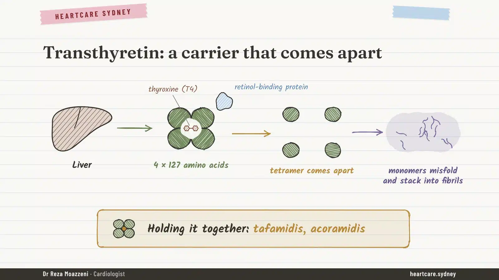
Transthyretin (the “TTR” in ATTR) is a normal blood protein made mainly by the liver. Its job is to carry thyroid hormone and vitamin A around the body. Four identical pieces lock together to form a stable unit called a tetramer.
The trouble starts when that unit comes apart. The single pieces fold the wrong way, stick together and build up as amyloid fibres, most of all in the heart. The heart walls thicken and stiffen, and the heart can no longer relax and fill well.
One disease, two types
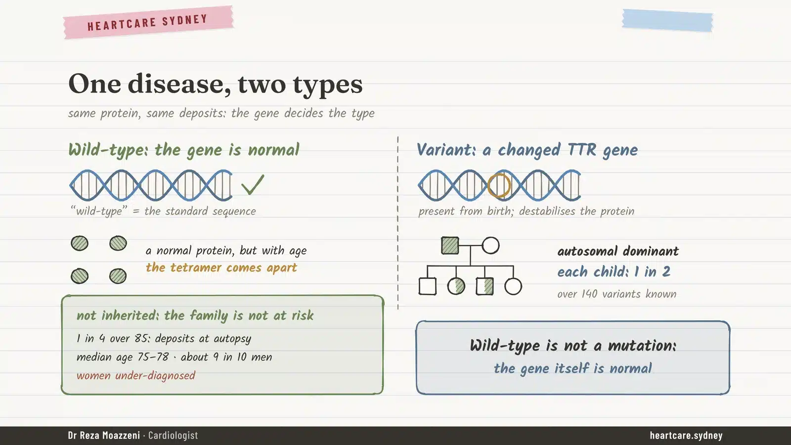
This is the part most people find confusing. There is one disease, ATTR amyloidosis, but two types, and the difference is the gene.
Wild-type ATTR: the gene is normal
“Wild-type” is a genetics term for the normal, standard version of a gene. In wild-type ATTR there is no mutation at all: the gene and the protein are completely normal. With age, the normal protein simply becomes less stable and starts to come apart. So wild-type ATTR is not inherited, and your family is not at extra risk.
It’s a condition of later life. At autopsy, about 1 in 4 people over 85 have some deposits. In clinics, patients are typically in their mid to late seventies, and about 9 in 10 are men, although women are probably under-diagnosed.
Hereditary (variant) ATTR: a changed gene
In hereditary ATTR, a change (a “variant”) in the TTR gene is present from birth and makes the protein less stable. It’s passed on in a dominant pattern: each child of a person with the variant has a 1 in 2 chance of inheriting it. More than 140 variants are known.
Wild-type is not a mutation: the gene itself is normal. Hereditary means a changed gene, and the family may be at risk.
The variant decides which organs are hit
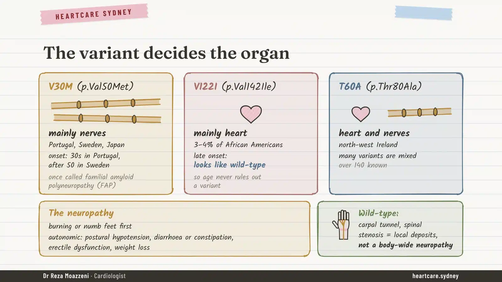
Where the amyloid settles depends on which variant a family carries. A few examples:
- V30M, common in Portugal, Sweden and Japan, mainly affects the nerves. It’s the condition once called familial amyloid polyneuropathy, often starting in the thirties in Portuguese families and after 50 in Swedish ones.
- V122I, carried by about 3 to 4% of African Americans, mainly affects the heart, late in life. It looks just like wild-type disease, which is why age alone never rules out a hereditary form.
- T60A, from north-west Ireland, affects both heart and nerves, as many variants do.
When the nerves are involved, the first sign is usually burning or numbness in the feet. The automatic nerves can be affected too, causing light-headedness on standing, diarrhoea or constipation, erectile problems and weight loss.
One point that often causes confusion: people with wild-type ATTR commonly have carpal tunnel syndrome or narrowing of the spinal canal. Those come from local amyloid deposits pressing on a nerve, not from a body-wide neuropathy.
Who needs a gene test, and how is it done?
Everyone diagnosed with ATTR should have a gene test, whatever their age. You can’t tell wild-type from hereditary disease by looking at the heart, and the answer changes three things:
- The nerves: a variant means checking for neuropathy, and it can open up treatments first approved for hereditary disease.
- The outlook: some variants, such as V122I, carry a worse prognosis.
- The family: relatives can find out whether they carry the variant.
Testing starts with genetic counselling, because a result affects the whole family. In Australia, the test is ordered by a specialist, with written consent, and is Medicare-funded for eligible patients. If a variant is found, first-degree relatives (parents, brothers and sisters, children) are offered counselling and testing for that specific variant from age 18. Carriers aren’t left waiting for symptoms: checks such as an ECG, Holter monitor, echocardiogram, NT-proBNP blood test and a nerve review usually start about 10 years before the age the disease typically begins in their family.
The Medicare neuromuscular gene panel (MBS 73422) includes TTR and requires signs of a neuromuscular disorder; relatives are then tested for the family variant (MBS 73423). The cardiomyopathy gene panel (MBS 73392) doesn’t list TTR, so ask the laboratory to include it. Counselling and written consent are expected before testing.
Where ATTR hides: the warning signs
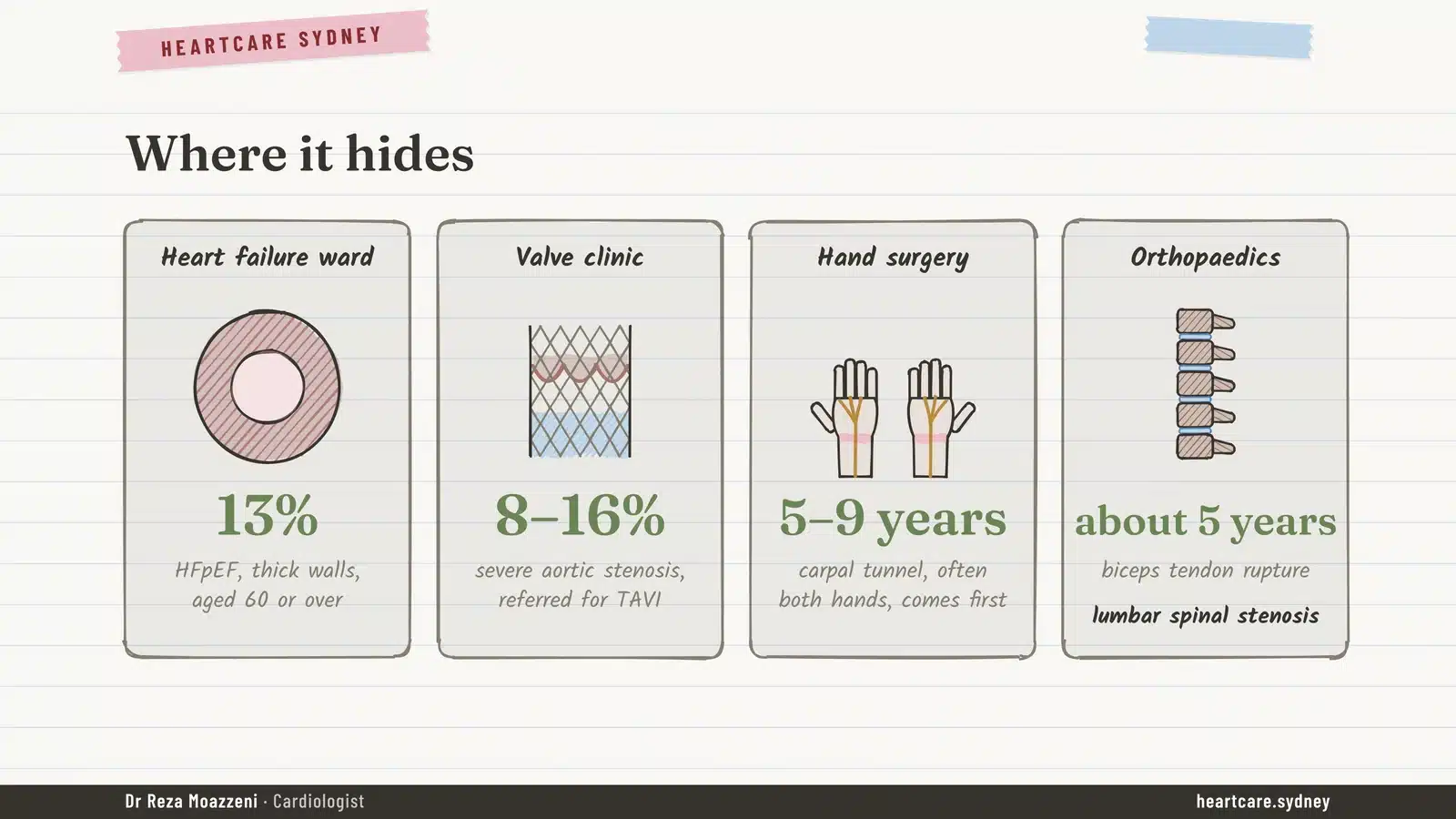
ATTR is still found late. In one large UK series, 42% of people with wild-type disease waited more than four years after their first heart symptoms before the diagnosis was made. These are the places it tends to turn up:
- About 13% of older people admitted with heart failure, normal pumping function and thick heart walls have ATTR.
- So do 8 to 16% of older people with severe aortic stenosis referred for TAVI.
- Carpal tunnel syndrome, often in both hands, comes 5 to 9 years before the heart diagnosis, and a ruptured biceps tendon at the elbow about 5 years before.
- Lumbar spinal stenosis is another clue.
The heart itself gives hints too. Because the stiff heart pumps a small volume with each beat, blood pressure often falls, and long-standing high blood pressure may seem to “get better” on its own. Atrial fibrillation is present in more than half of people with wild-type ATTR, and heart blood tests (troponin and NT-proBNP) are often higher than expected. The classic “low voltage” ECG appears in only about 1 in 5.
How is it diagnosed?
Echocardiogram and MRI
An echocardiogram usually shows walls 12 mm or thicker, often with a thick right ventricle, thick valves, enlarged upper chambers and a small amount of fluid around the heart. A special analysis of heart muscle motion (strain) often shows the bottom of the heart squeezing better than the base. Cardiac MRI adds a characteristic pattern of scarring. These tests suggest amyloid, but neither can tell ATTR from AL.
The PYP scan and blood tests, done together
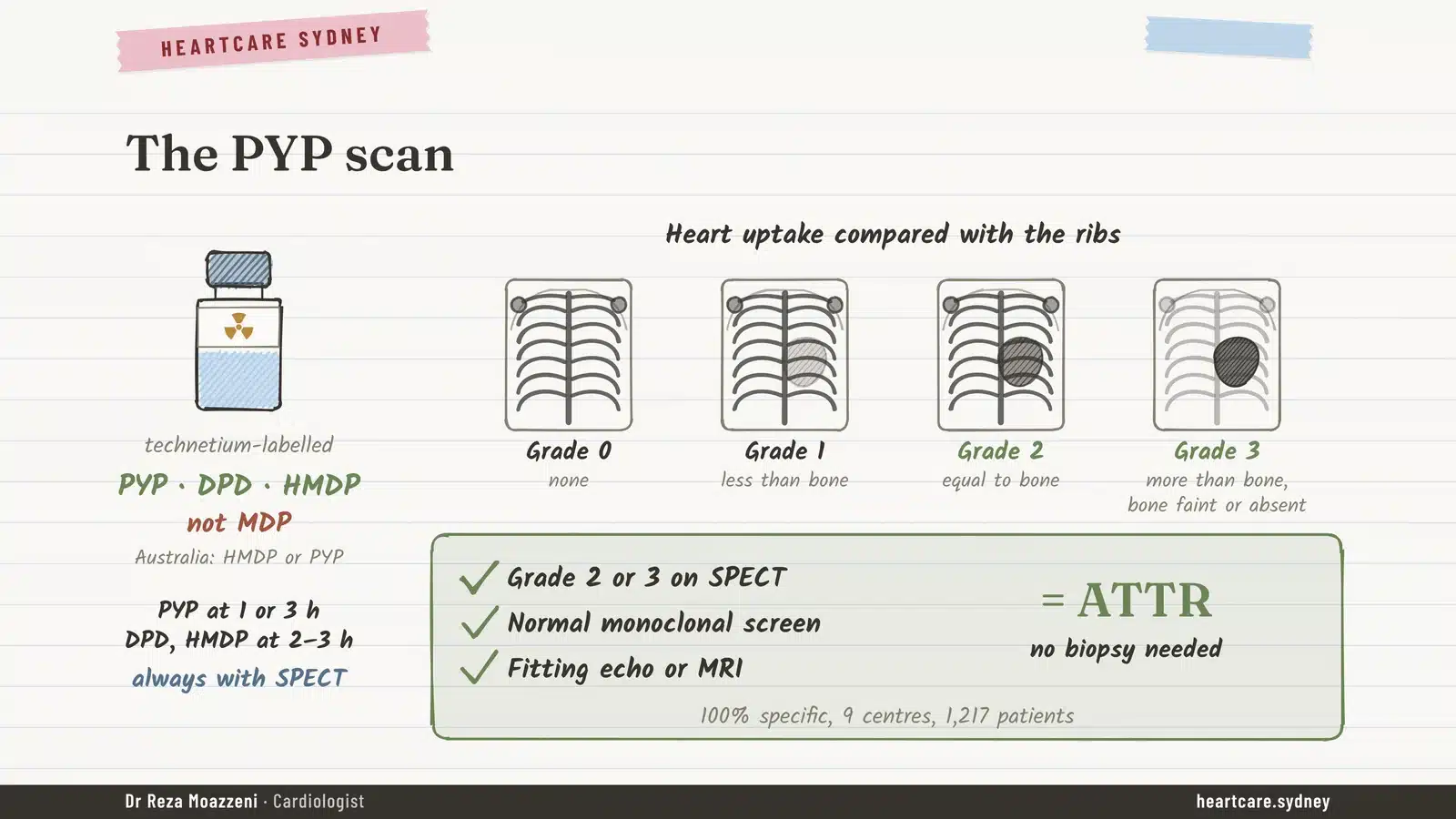
The key test is a nuclear heart scan using a bone tracer, called a PYP scan (similar tracers are DPD and HMDP). ATTR deposits soak up the tracer. The scan is always combined with SPECT, a 3D view that makes sure the signal is truly in the heart muscle and not just blood in the heart chambers.
Uptake is graded from 0 to 3 by comparing the heart with the ribs, a method called the Perugini score:
- Grade 0: no heart uptake. ATTR is unlikely, though not impossible.
- Grade 1: the heart is fainter than the ribs. This is equivocal and needs more tests.
- Grade 2: the heart is about as bright as the ribs.
- Grade 3: the heart is brighter than the ribs, and the bones look faint.
The key point: a grade 2 or 3 scan, together with normal blood and urine tests for the AL protein and a fitting echo or MRI, diagnoses ATTR without a biopsy. Across nine specialist centres, that combination was 100% specific. That’s why the blood and urine tests are done at the same time as the scan, never skipped.
When the scan can mislead
On its own, the scan isn’t enough. About 4 in 10 people with AL amyloidosis show some heart uptake, and a few reach grade 2 or 3. Some hereditary variants can give a negative scan despite real disease. A grade 1 result, or a negative scan when suspicion remains high, calls for MRI, specialist review or a biopsy. A fat pad biopsy, so useful in AL, finds only about 15% of wild-type ATTR.
Putting it together: the diagnostic pathway
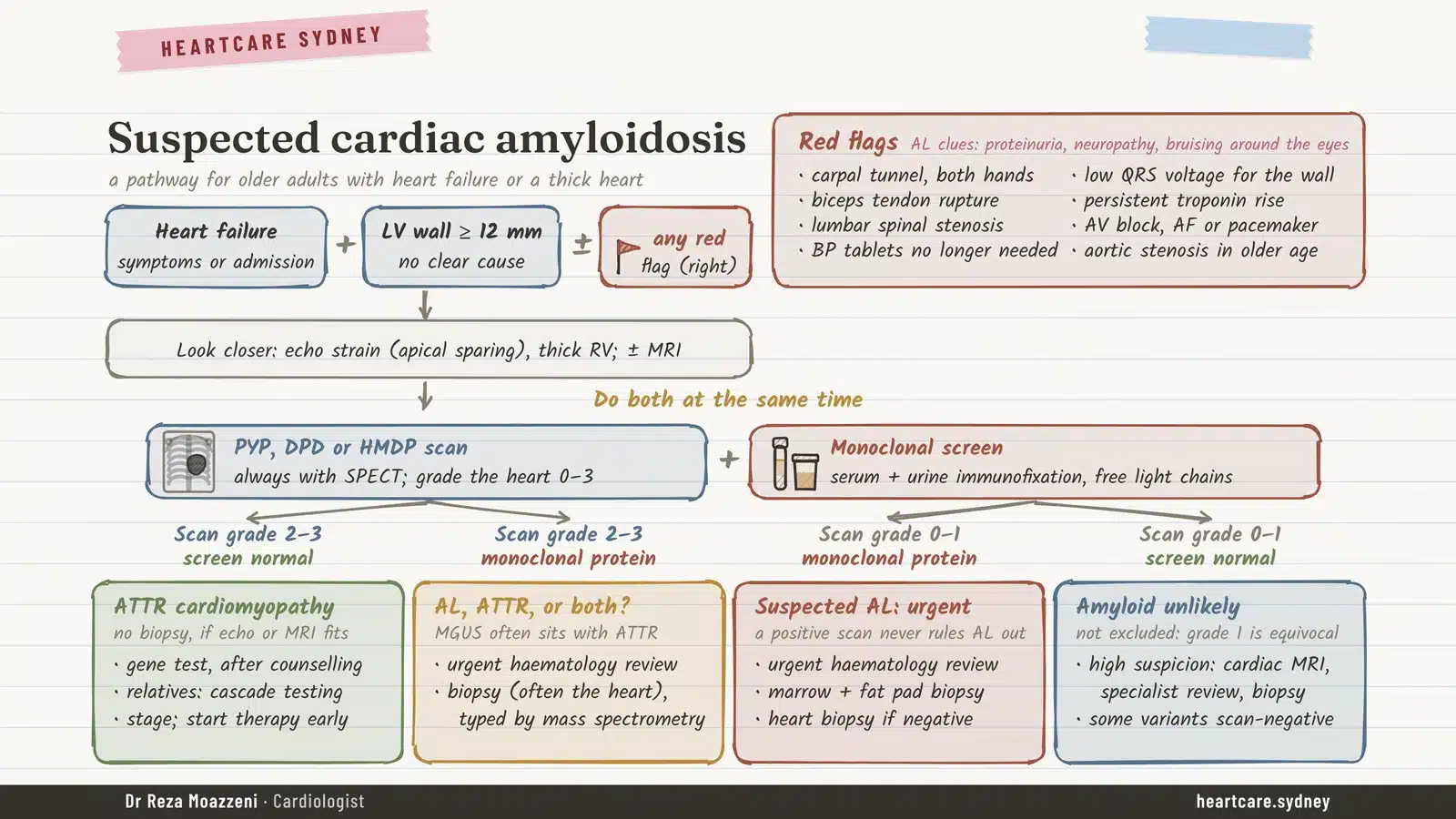
The chart below brings these steps together. It starts with heart failure or a thick heart with no clear cause, and the red flags that should prompt a closer look. The scan and the blood and urine tests are then done at the same time, and the combination of results points to one of four next steps.
How serious is it?
ATTR is slower than AL but still serious. In the UK study that set up the staging system, median survival was 69 months when two blood tests (NT-proBNP and kidney function) were both reassuring, 47 months with one abnormal and 24 months with both. Those patients were diagnosed before modern drugs, and survival has improved since: at one centre, median survival rose from about 3 years in the mid-2000s to more than 5 years by 2017 to 2021, partly because the disease is now found earlier.
Treatment
Looking after the heart
Some of the usual heart failure treatments suit ATTR and some don’t:
- Fluid tablets (diuretics) are the mainstay, but the window is narrow, because these stiff hearts depend on being well filled.
- Spironolactone and SGLT2 inhibitors are well tolerated and linked to lower mortality in observational studies.
- Beta-blockers and ACE inhibitors are often poorly tolerated. In one large series, only about 6% of patients reached full beta-blocker doses, with no clear benefit unless the pumping function was reduced.
- Verapamil and diltiazem should be avoided. Digoxin can be used cautiously at a low dose.
- Atrial fibrillation needs a blood thinner whatever the usual risk score. Amiodarone is the preferred rhythm medicine.
- Some people need a pacemaker. Defibrillators haven’t been shown to help people live longer in ATTR.
- Severe aortic stenosis deserves a referral to the valve team: in one study, one-year mortality was 16% after valve replacement versus 55% without.
Tafamidis: holding the protein together
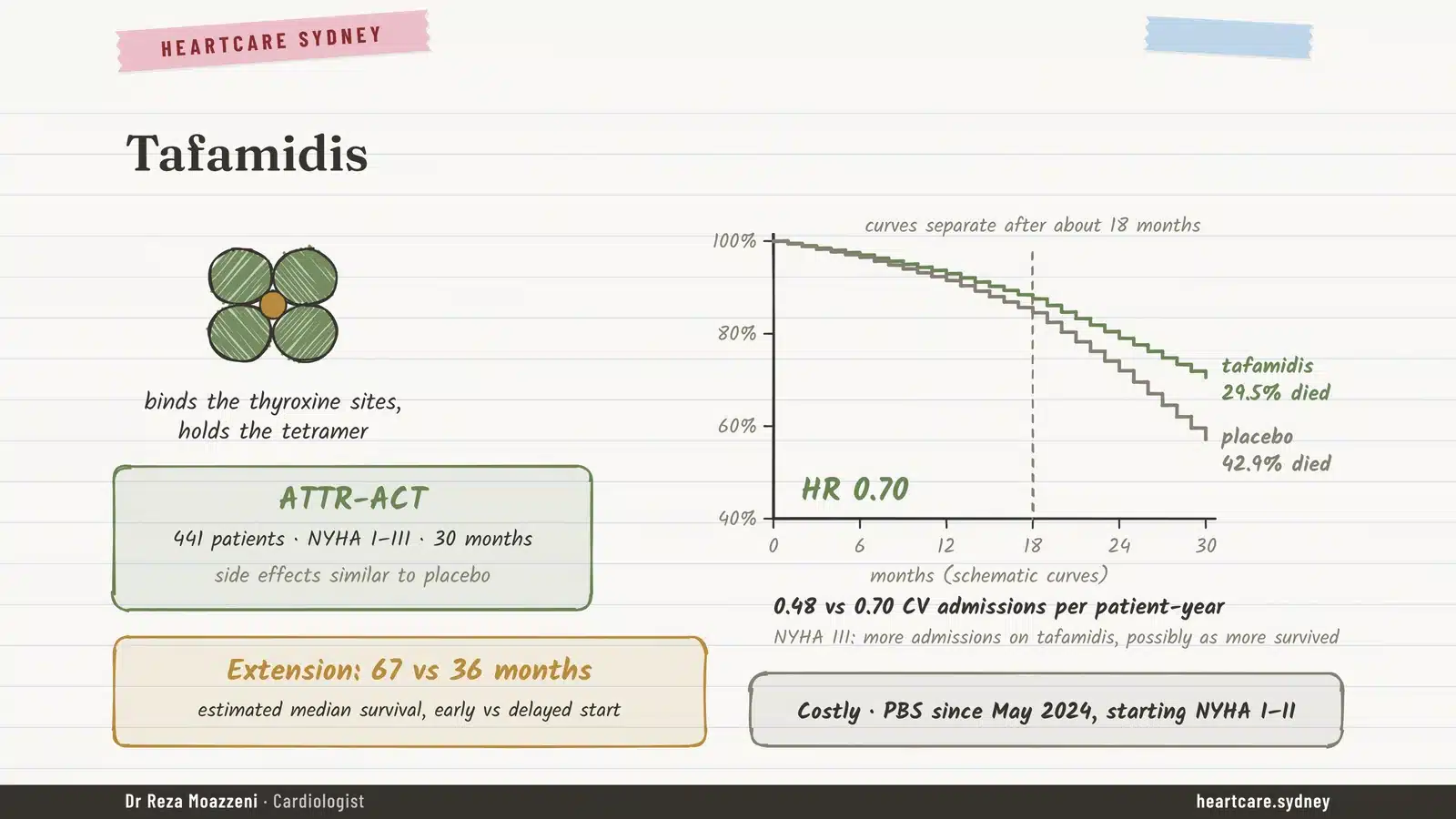
Tafamidis binds to transthyretin and keeps the four pieces locked together, so less amyloid forms. In the ATTR-ACT trial, 441 people took tafamidis or a placebo for 30 months. Deaths fell from 43% to 30%, heart-related hospital admissions fell by about a third, and side effects were similar to placebo.
The benefit takes time to show (the survival curves only separated after about 18 months), and starting early matters. In the trial’s extension, people who started tafamidis from the beginning had an estimated median survival of 67 months, compared with 36 months for those who started later. In Australia, tafamidis is PBS-listed for people who start it while their symptoms are mild (NYHA class 1 or 2).
Newer medicines
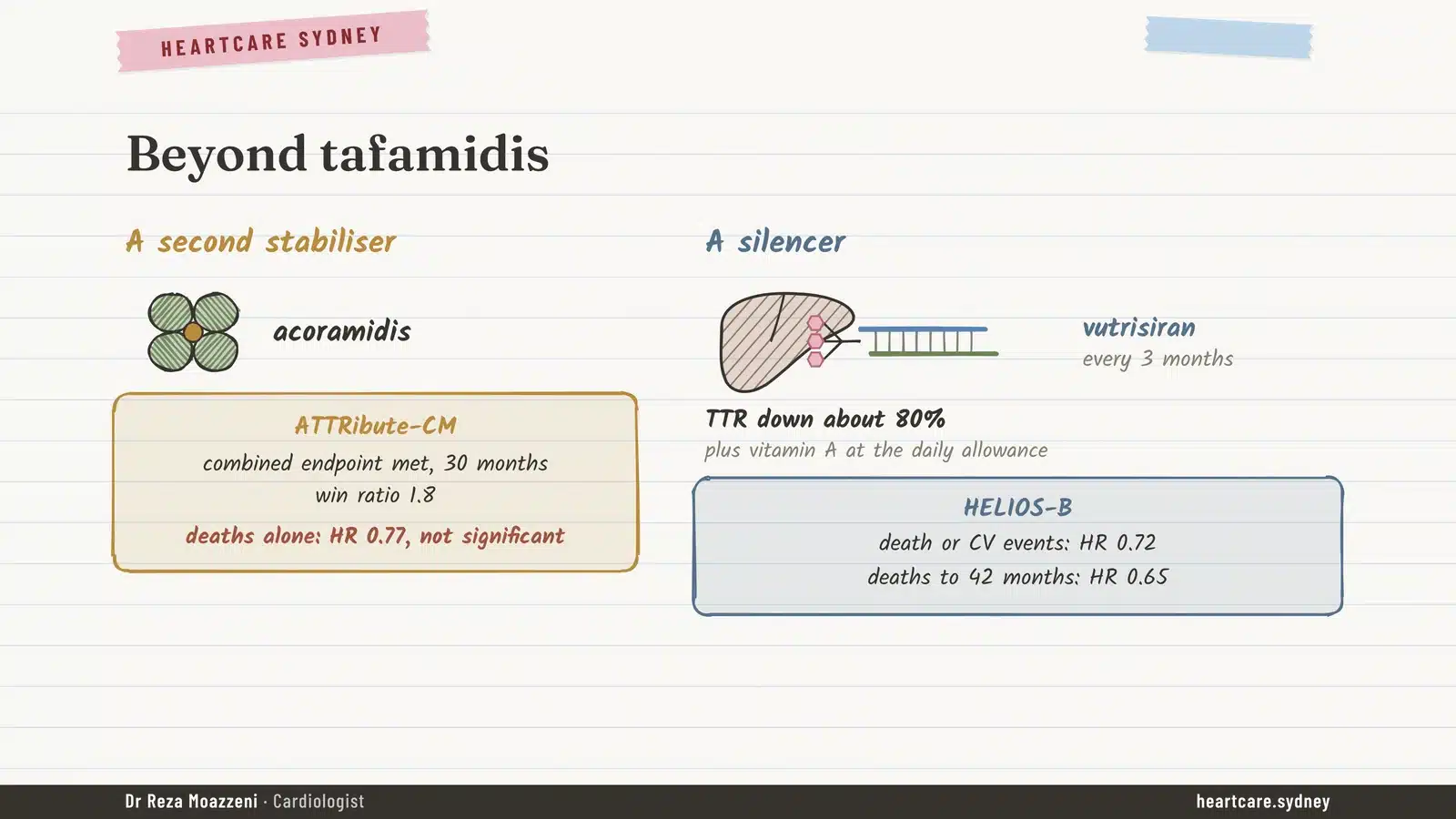
Acoramidis is a second stabiliser that works like tafamidis. In the ATTRibute-CM trial it met its main combined goal of fewer deaths and hospital admissions, although the effect on deaths alone wasn’t statistically significant.
Vutrisiran works differently. It’s a “silencer”: an injection every three months that tells the liver to make about 80% less transthyretin. In the HELIOS-B trial it reduced death or heart events by about a quarter, and deaths by about a third. Because transthyretin also carries vitamin A, people on vutrisiran take a vitamin A supplement at the usual daily allowance.
Not every silencer has worked in the heart. A related drug, eplontersen, did not reduce heart deaths and events overall in the CARDIO-TTRansform trial, reported in 2026. Whether combining two drugs (say, a stabiliser plus a silencer) adds benefit is still unproven, so the choice rests on the trial evidence, access and cost.
In Australia, the PBS advisory committee recommended vutrisiran in July 2026 for people with mild symptoms (NYHA class 1 or 2), but it isn’t listed yet, and acoramidis is due for consideration in November 2026.
- ATTR is one disease with two types: wild-type (normal gene, age-related, not inherited) and hereditary (a changed gene, 1 in 2 chance for each child).
- Everyone with ATTR should have a gene test after counselling, and relatives of a carrier should be offered testing.
- Think of ATTR in older people with thick heart walls and heart failure, especially with carpal tunnel syndrome or aortic stenosis.
- A grade 2 or 3 PYP scan plus normal AL blood and urine tests diagnoses ATTR without a biopsy.
- Tafamidis works best started early; a second stabiliser and a silencer have also shown benefit in trials.
How our clinic can help
At Heartcare Sydney we see many people with thick heart walls, heart failure with normal pumping function, atrial fibrillation or aortic stenosis, the very groups in which ATTR hides. If amyloid is possible, we assess the heart in detail with echocardiography, arrange the PYP scan and the blood and urine tests together, and guide gene testing, family screening and treatment. If you’ve had carpal tunnel surgery on both hands and now have heart symptoms, it’s worth mentioning to your doctor.
The other main type of heart amyloid, AL, starts in the bone marrow and needs urgent treatment. Read about it in our article on AL amyloidosis and the heart.
Thick heart walls, unexplained heart failure, or carpal tunnel on both sides?
An assessment, including an echocardiogram, can help work out whether amyloid is the cause. A GP referral is required.
Book an appointmentFrequently asked questions
Is ATTR amyloidosis inherited?
Only the hereditary (variant) type. Wild-type ATTR, the more common type in older people, comes from a normal gene and isn’t passed on. A gene test tells the two apart, which is why everyone with ATTR should have one.
What does “wild-type” mean?
It’s the genetics term for the normal, standard version of a gene. Wild-type ATTR means the transthyretin gene is normal; the protein becomes unstable with age rather than because of a mutation.
Do I need a heart biopsy to diagnose ATTR?
Usually not. A grade 2 or 3 PYP scan with SPECT, normal blood and urine tests for the AL protein, and a fitting echo or MRI together diagnose ATTR without a biopsy.
Should my children be tested?
Only if you carry a hereditary variant. Then each child has a 1 in 2 chance of carrying it, and adult relatives are offered genetic counselling and testing, usually from 18. Carriers start regular heart and nerve checks about 10 years before the age the disease usually begins in the family.
Can ATTR be cured?
Not yet. Current treatments slow or stop new amyloid forming: stabilisers such as tafamidis and acoramidis hold the protein together, and silencers such as vutrisiran reduce how much is made. They work best when started early.
References
Main sources
- Maurer MS, et al. Tafamidis treatment for patients with transthyretin amyloid cardiomyopathy (ATTR-ACT). N Engl J Med. 2018.
- Gillmore JD, et al. Acoramidis in transthyretin amyloid cardiomyopathy (ATTRibute-CM). N Engl J Med. 2024.
- Fontana M, et al. Vutrisiran in patients with transthyretin amyloidosis with cardiomyopathy (HELIOS-B). N Engl J Med. 2025.
- Gillmore JD, et al. Nonbiopsy diagnosis of cardiac transthyretin amyloidosis. Circulation. 2016.
- Perugini E, et al. Noninvasive etiologic diagnosis of cardiac amyloidosis using 99mTc-DPD scintigraphy. J Am Coll Cardiol. 2005.
- Pharmaceutical Benefits Scheme (PBS) records as at 1 November 2026.
Figures come from published studies and guidelines current in November 2026. The illustrations are schematic and show no real patient results. Please don’t start, stop or change any medicine without talking to your doctor.