
Sometimes a heart scan shows thick heart walls in someone who has never had high blood pressure. Usually there is a simple explanation. Occasionally, the reason is amyloid: an abnormal protein that has built up inside the heart muscle. One of the two main types, AL amyloidosis, starts in the bone marrow, moves quickly and responds best to treatment when it’s found early. This article explains what it is, how we find it and how it’s treated today.
What is AL amyloidosis?
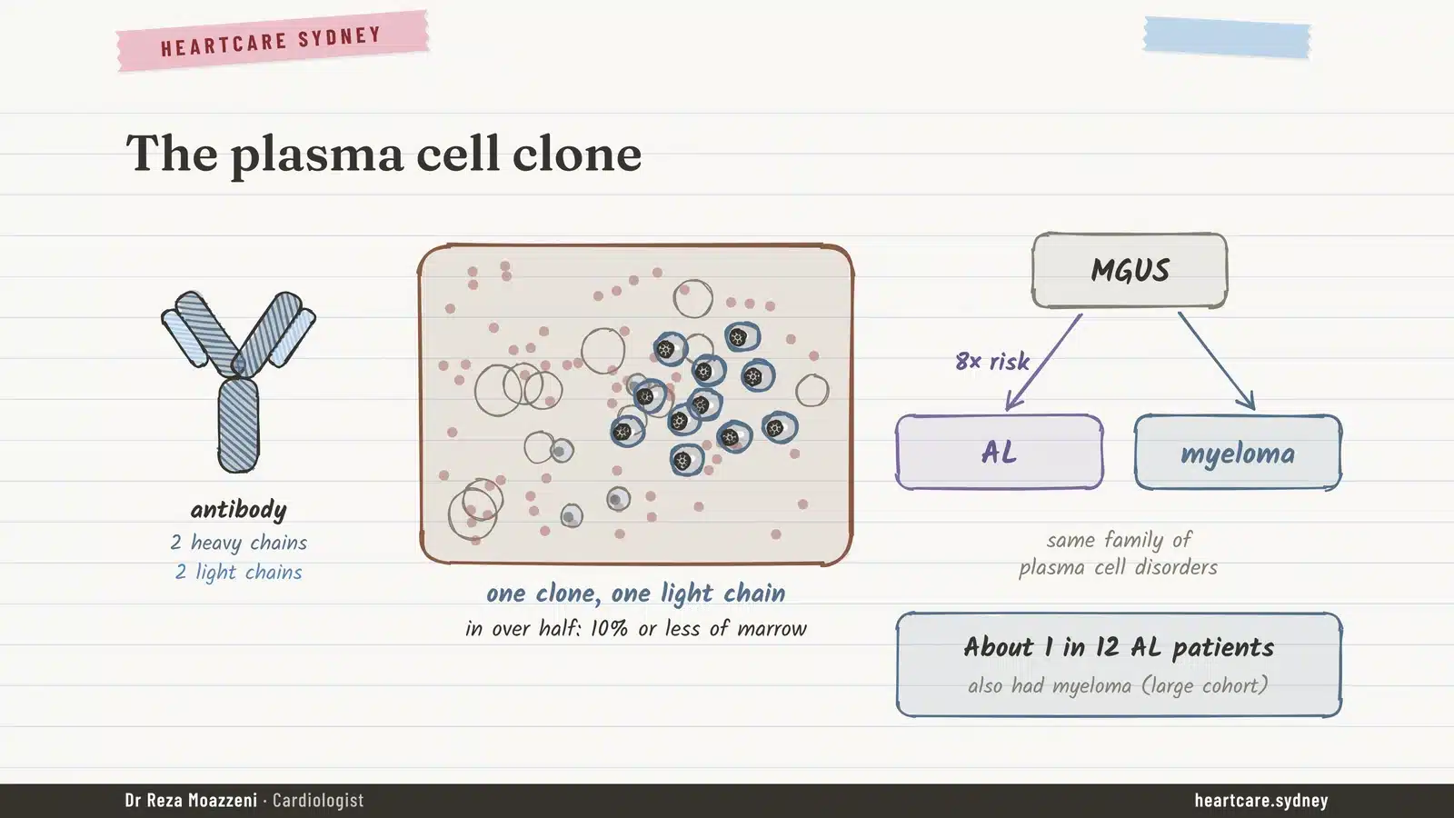
Plasma cells live in the bone marrow and make antibodies. Each antibody is built from two “heavy” and two “light” protein chains. Normally the marrow holds many different plasma cells, each making its own antibody, and a small surplus of light chains spills into the blood, where the kidneys clear it within hours.
In AL amyloidosis, one plasma cell multiplies into a family of identical cells, called a clone. That clone makes large amounts of a single abnormal light chain. Some of these light chains are unstable: they unfold, stick together and form tiny fibres called amyloid, which settle in organs. “AL” simply stands for amyloid made of light chains.
The clone is usually small. In more than half of patients, plasma cells make up 10% or less of the marrow, which is why AL is not the same as myeloma, although it belongs to the same family of blood cell conditions. People with a common, harmless-looking finding called MGUS (a “monoclonal gammopathy of undetermined significance”) have about eight times the usual risk of developing AL, and about 1 in 12 people with AL also have myeloma.
AL is rare: about 10 to 12 new cases per million people each year. But it moves fast. Without treatment, people who already have heart failure from AL live a median of under six months. That’s why we treat any suspicion of AL as urgent.
How does it harm the heart?
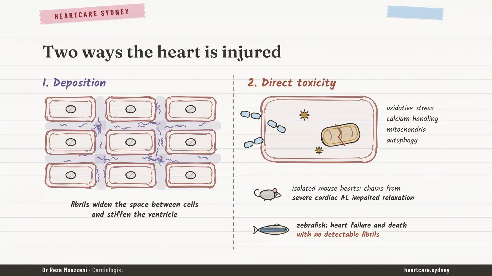
The light chains damage the heart in two ways.
- Deposits. Amyloid fibres build up between the heart muscle cells. The walls thicken and stiffen, so the heart can’t relax and fill properly. Think of trying to fill a thick rubber balloon instead of a thin one.
- Direct toxicity. The circulating light chains are toxic to heart cells in their own right, even before many fibres have formed. In laboratory studies, light chains from patients with severe heart involvement stopped heart muscle relaxing properly and harmed the cells’ energy supply, and in zebrafish they caused heart failure with no visible deposits at all.
We see the same thing in patients. When chemotherapy cuts the light chains by more than half within three cycles, a heart strain marker in the blood (NT-proBNP) falls by about half, even though the heart walls look no thinner yet. When the light chains don’t fall, NT-proBNP rises, and those patients account for most deaths. In practice, this means switching off the light chains quickly protects the heart, sometimes within weeks.
It’s rarely just the heart
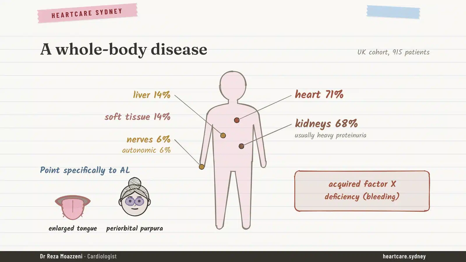
AL is a whole-body disease. In a large UK group of 915 patients, the heart was involved at diagnosis in 71% and the kidneys in 68%, usually with heavy protein loss in the urine. The liver and soft tissues were each involved in about 1 in 7, and the nerves in about 1 in 16.
A few signs point specifically to AL:
- an enlarged tongue, sometimes with tooth marks along its edges
- bruising around the eyes after minor strain, such as coughing
- easy bleeding from a lack of a clotting factor (factor X), which the amyloid soaks up
Symptoms that should make us think of it
Early symptoms are vague, which is why AL is often found late. The ones we listen for:
- breathlessness, ankle swelling or tiredness that doesn’t fit the usual explanations
- a heart scan showing thick walls without high blood pressure or a valve problem
- blood pressure that has fallen, so tablets that used to suit now cause dizziness
- frothy urine or swelling from protein loss through the kidneys
- numbness or burning in the feet, light-headedness on standing, or bowel changes
- weight loss, an enlarged tongue or easy bruising around the eyes
How is AL diagnosed?
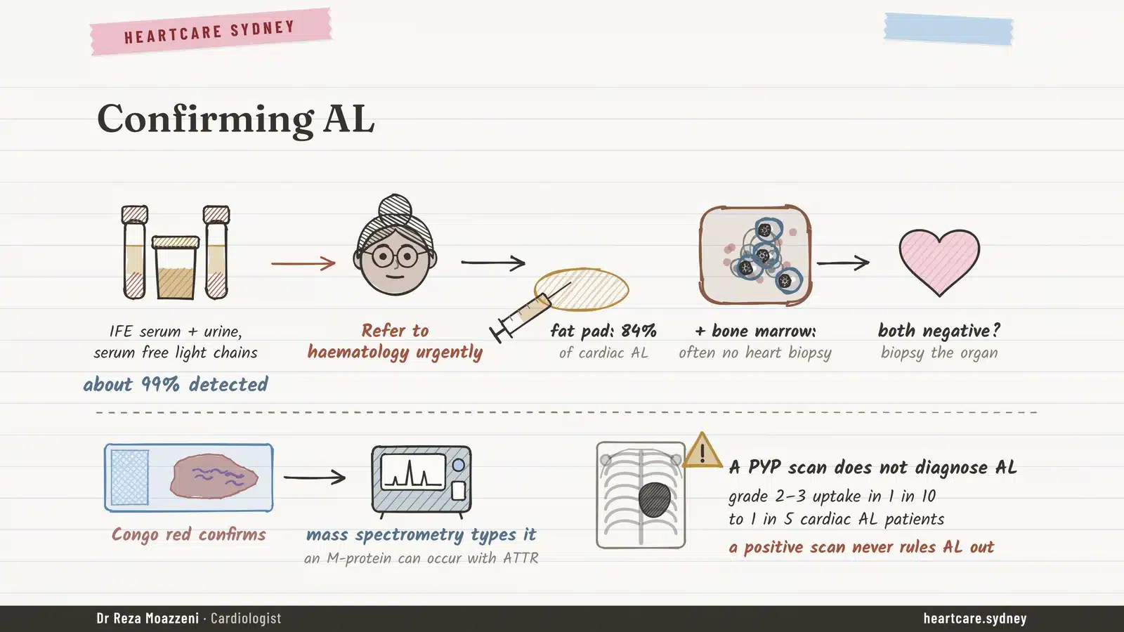
The first step is a set of blood and urine tests: serum free light chains plus immunofixation of both blood and urine. Together they find the abnormal protein in about 99% of people with AL. A simple protein electrophoresis on its own isn’t enough.
If any of these tests is abnormal, the next steps happen in parallel, not one after another:
- Urgent referral to a haematologist, who treats the clone.
- A tissue sample to prove amyloid is present. A small sample of fat from the tummy wall is positive in about 84% of people with heart involvement, and together with a bone marrow biopsy it often avoids the need for a heart biopsy.
- In the laboratory, a dye called Congo red confirms amyloid, and a test called mass spectrometry shows exactly which protein it is made of.
That last step matters because the other main type of heart amyloid, ATTR, is common in older people, and so are harmless abnormal proteins in the blood. A person can have both. The bone-tracer heart scan (a PYP scan) is excellent for ATTR but cannot diagnose AL: most AL hearts take up little tracer, yet up to 1 in 5 light up just like ATTR. A positive scan never rules AL out.
How serious is it? The heart sets the pace
In AL, the heart decides the outlook more than any other organ. Doctors stage the disease with three blood tests: two heart markers (troponin and NT-proBNP) and the size of the abnormal light chain excess. Before modern treatment, people with none of these raised lived a median of about eight years; people with all three raised, about six months. The sickest group, with very high NT-proBNP, still does poorly today, which is the strongest argument for finding AL earlier.
Treatment: switching off the clone
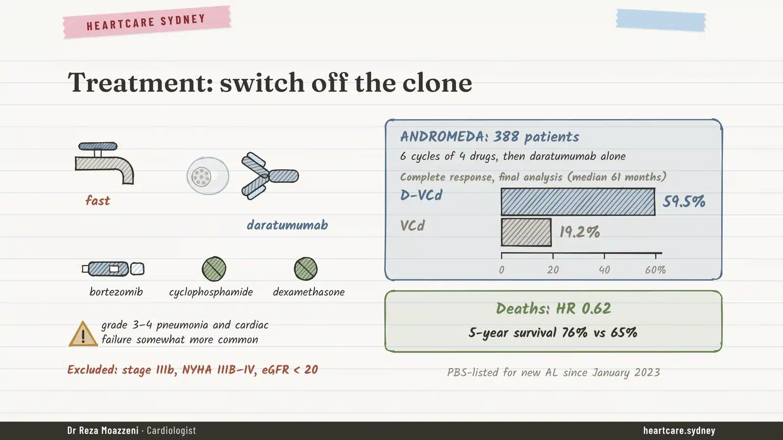
Treatment aims to switch off the plasma cell clone as quickly and as deeply as possible, because every week its light chains keep harming the heart. The haematologist leads this part of care.
The standard first treatment combines an antibody, daratumumab, with three older drugs: bortezomib, cyclophosphamide and dexamethasone. In the ANDROMEDA trial, adding daratumumab raised the rate of complete blood response from 19% to about 60%, and five years on, 76% of patients were alive compared with 65%. Chest infections and heart failure were somewhat more common, so these patients need close monitoring. In Australia, daratumumab has been on the PBS for newly diagnosed AL since January 2023.
Some fitter patients, usually under 70 and without advanced heart involvement, can have high-dose chemotherapy with a stem cell transplant. Its early risk has fallen a lot: deaths within 100 days dropped from about 20% to about 5%. Antibodies designed to dissolve the amyloid itself have, so far, not worked in large trials, so treating the clone remains the cornerstone.
How do we know it’s working?
Response comes in two steps. The blood responds first. Within a few cycles, the light chain excess should fall to a low level; if it hasn’t fallen enough after about three cycles, many centres change treatment.
The heart follows later, typically taking around a year to show its best improvement. It’s worth the wait: in one large study, 93% of people whose heart responded fully were alive at five years, compared with 33% of those whose heart didn’t respond. We track the heart with NT-proBNP, symptoms and imaging such as echocardiography.
Looking after the heart along the way
While the haematology treatment does its work, our job is to keep the heart and the patient well enough to get through it. A few things are different from ordinary heart failure:
- Fluid tablets (diuretics) relieve swelling and breathlessness, but stiff hearts depend on having enough filling, so we use them gently and watch blood pressure and kidney function.
- Spironolactone and SGLT2 inhibitors may help and are usually well tolerated. The best evidence comes from the other type, ATTR; in AL, the data are still thin.
- Beta-blockers and ACE inhibitors, staples of other heart failure, are often poorly tolerated, because the heart relies on its rate to keep output up and blood pressure is often low.
- Digoxin is used only with caution.
- Blood thinners are recommended for atrial fibrillation whatever a person’s usual risk score, because clots in the heart are common in AL.
- Defibrillators can deliver shocks, but they haven’t been shown to help people live longer in AL, so the decision is individual.
The outlook today
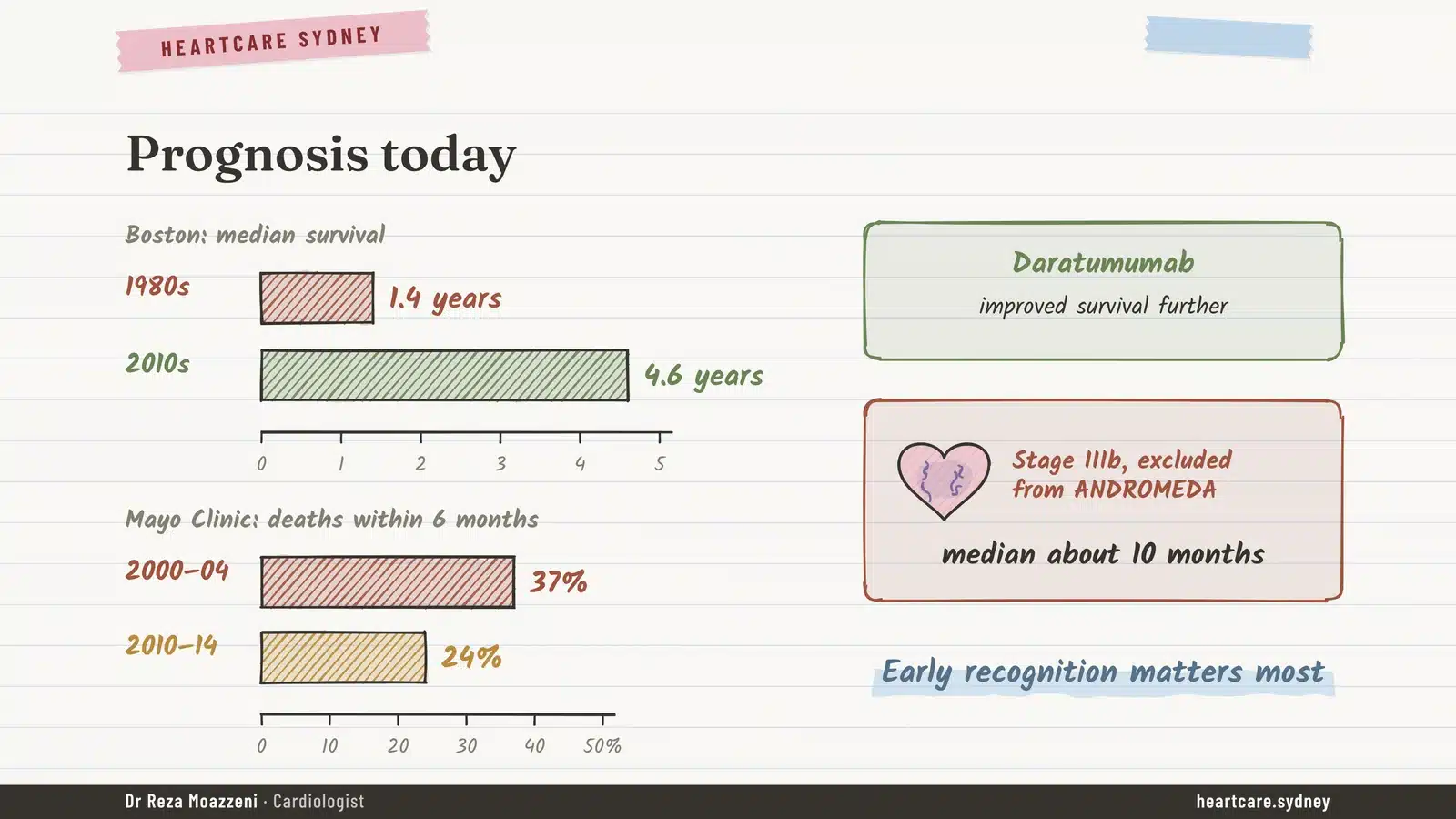
The picture has changed a great deal. At one large US centre, median survival rose from 1.4 years for people diagnosed in the 1980s to 4.6 years for those diagnosed in the 2010s, and that was before daratumumab became standard. Early deaths have fallen too.
That last figure is why early diagnosis matters so much. The biggest gains come from finding AL before the heart is badly damaged.
- AL amyloidosis is a bone marrow plasma cell disorder whose light chains damage the heart in two ways: deposits and direct toxicity.
- It’s rare but fast-moving, and the heart decides the outlook.
- The screening tests are simple: serum free light chains plus blood and urine immunofixation.
- An abnormal result means urgent haematology referral and a tissue sample at the same time.
- Treatment with daratumumab has improved results; the blood responds in weeks to months, the heart over about a year.
How our clinic can help
At Heartcare Sydney we see people with unexplained thick heart walls, heart failure with a normal pumping function, or symptoms that don’t add up. If amyloid is a possibility, we check the heart in detail with echocardiography, arrange the blood and urine tests, and work alongside your haematology team so the heart is looked after while the clone is treated. If AL turns out not to be the cause, we’ll look for what is.
The other main type of heart amyloid, ATTR, behaves very differently. You can read about it in our article on ATTR amyloidosis and the heart.
Been told your heart walls are thick?
If you have unexplained breathlessness, swelling or a scan showing thick heart walls, an assessment with an echocardiogram can help find the cause. A GP referral is required.
Book an appointmentFrequently asked questions
Is AL amyloidosis a type of cancer?
It’s a disorder of plasma cells, which are blood cells in the bone marrow, and it belongs to the same family as myeloma. The clone is usually small, so most people with AL don’t have myeloma, but it’s treated by haematologists with similar drugs.
Is AL amyloidosis inherited?
No. AL comes from a clone of plasma cells that develops during life. It isn’t passed on to children. (Some forms of the other type, ATTR, can be inherited.)
What tests check for AL amyloidosis?
Serum free light chains plus immunofixation of the blood and urine. Together they find the abnormal protein in about 99% of people with AL. If anything is abnormal, a haematologist and a tissue sample confirm the diagnosis.
Can a heart scan diagnose AL?
An echocardiogram or cardiac MRI can show the pattern of amyloid, but not which type it is. The bone-tracer (PYP) scan identifies ATTR, not AL, and up to 1 in 5 AL hearts can light up on it. AL is confirmed with blood tests and a tissue sample.
Can the heart recover?
Often, yes. Once the light chains are switched off, heart markers can improve within weeks to months, and the heart usually reaches its best response after about a year. People whose heart responds fully do much better over the long term.
References
Main sources
- Kastritis E, et al. Daratumumab-based treatment for immunoglobulin light-chain amyloidosis (ANDROMEDA). N Engl J Med. 2021.
- UK and US amyloidosis cohort studies quoted in the text (organ involvement, staging, survival trends).
Figures come from published studies and guidelines current in November 2026. The illustrations are schematic and show no real patient results. Please don’t start, stop or change any medicine without talking to your doctor.