
Hypertrophic cardiomyopathy (HCM) can cause severe left ventricular outflow tract obstruction (LVOTO) without striking hypertrophy. These two patients had a maximum basal septal thickness of 15 mm and normal overall LV mass, yet both developed marked resting obstruction. The older patient reached a peak instantaneous gradient of approximately 256 mmHg with Valsalva.
Both patients consented to the use of de-identified investigations for education. Some details have been altered to protect privacy.
- Why these cases matter
- Case 1: from 7 to 94 mmHg at rest
- Case 2: above 200 mmHg with few reported symptoms
- LVOT jet or mitral regurgitation?
- HCM or hypertensive remodelling?
- The two cases at a glance
- Why modest hypertrophy can obstruct
- What cardiac MRI adds
- Genetic results and the family
- Sudden-death risk assessment
- Treatment
- Medications that worsen LVOTO
- Aficamten: ESC 2026 update
- Key takeaways
- References
A 15 mm septum can still cause severe obstruction
Both patients had earlier echocardiograms showing resting gradients of only 6–8 mmHg. A few years later, the younger patient presented with dyspnoea and presyncope on exertion. The other patient, who had a new murmur on cardiac auscultation, reported few symptoms during her predominantly sedentary routine, although exercise testing elicited dyspnoea. The earlier studies establish low resting gradients at those visits.
Metoprolol reduced resting gradients in both patients and markedly improved the younger patient’s symptoms. Valsalva continued to provoke severe obstruction. The cases illustrate how septal shape, mitral valve motion, contractility and loading conditions can matter as much as the wall-thickness measurement.
Case 1: from 7 to 94 mmHg at rest
A woman in her late 50s presented in 2025 with exertional dyspnoea and presyncope. Her background included well-controlled hypertension and type 2 diabetes. A CT coronary angiogram showed a calcium score of zero and no coronary artery disease.
In 2023, continuous-wave (CW) and pulsed-wave Doppler showed LVOT velocities of approximately 1.3–1.4 m/s, corresponding to resting peak gradients of 7–8 mmHg.
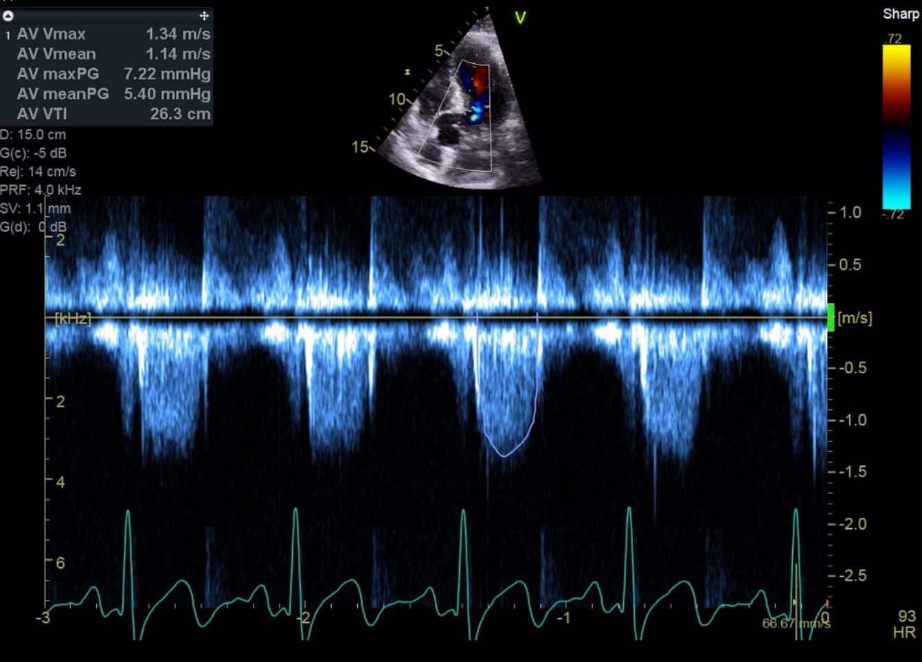
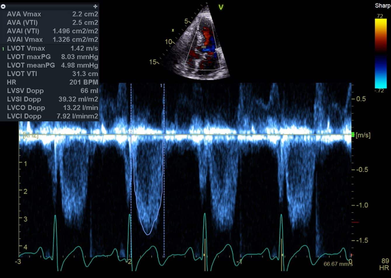
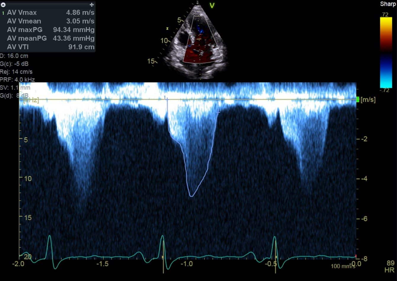
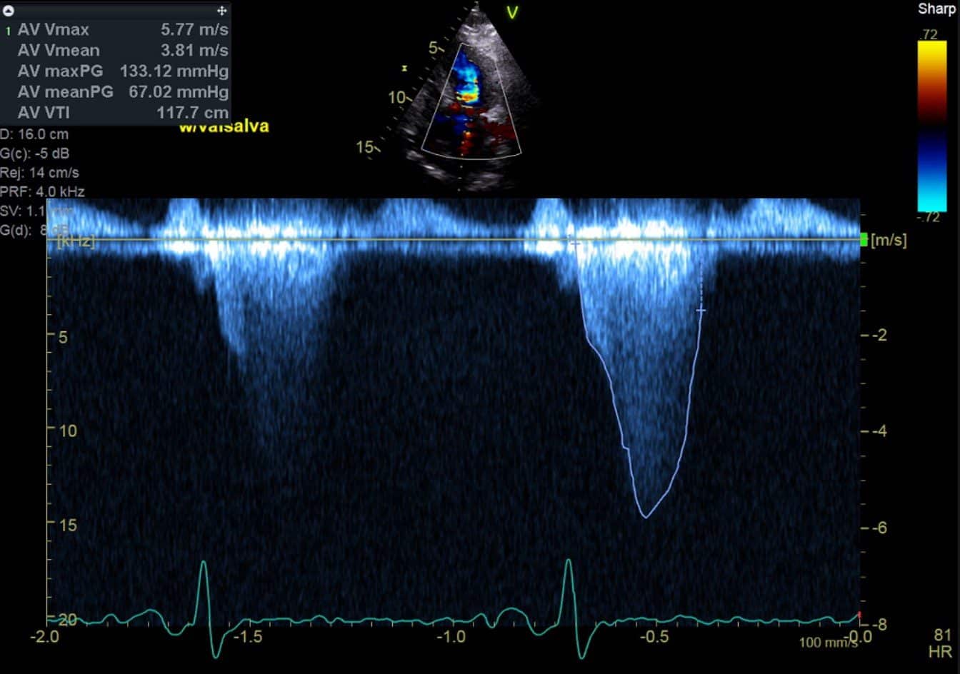
By 2025, severe obstruction was evident at rest. LVOT velocity was 4.86 m/s, giving a peak gradient of 94 mmHg. With Valsalva, this rose to 5.77 m/s and 133 mmHg. The late-peaking Doppler envelope and cine loops showed systolic anterior motion (SAM) of the mitral valve with marked narrowing of the LVOT.
The LV appeared relatively small and hyperdynamic on echocardiography. The degree of obstruction was striking in a ventricle without extensive hypertrophy.
Presentation cine loops
Cardiac MRI findings
Cardiac MRI showed a normal-sized LV, an ejection fraction of 69% and an indexed LV mass of 48 g/m². Both echocardiography and cardiac MRI showed focal basal anteroseptal thickening, with a maximum thickness of 15 mm. Echo-derived LV mass index was approximately 86 g/m², also within its reported reference range. CMR also described apicobasal muscle bands and LVOT turbulence, suggesting a possible contribution from accessory muscular structures.
Patchy mid-wall late gadolinium enhancement (LGE) involved the basal-to-mid anteroseptum and basal inferolateral wall. The report described a moderate burden, without a percentage of LV mass. This indicates myocardial fibrosis, but cannot be assumed to meet the threshold for extensive LGE used in HCM risk assessment. No myocardial oedema was reported.
Genetic testing: A TNNT2 variant of uncertain significance
Genetic testing identified a heterozygous TNNT2 variant, NM_001276345.2:c.341C>T (p.Ala114Val), also described as p.Ala104Val on another transcript. The testing laboratory classified it as a variant of uncertain significance (VUS). ClinVar contains conflicting classifications, so the result needs specialist interpretation and periodic review.4
Her brother reportedly has a similar phenotype, which raises the possibility of familial HCM. Testing an affected relative may help assess whether the variant tracks with disease in the family. Until its significance is resolved, however, this VUS should not be used to predict disease in unaffected relatives or to release them from clinical screening.1,2,4
Response to metoprolol
Metoprolol was started and titrated to 100 mg in the morning and 75 mg in the evening. Exertional dyspnoea and presyncope improved markedly. In 2026, the resting gradient was approximately 61 mmHg (3.90 m/s), while Valsalva still produced a gradient of approximately 124 mmHg (5.56 m/s).
Follow-up imaging showed persistent SAM and flow acceleration, with a less hyperdynamic resting appearance. Symptoms improved substantially despite little change in the provoked gradient.
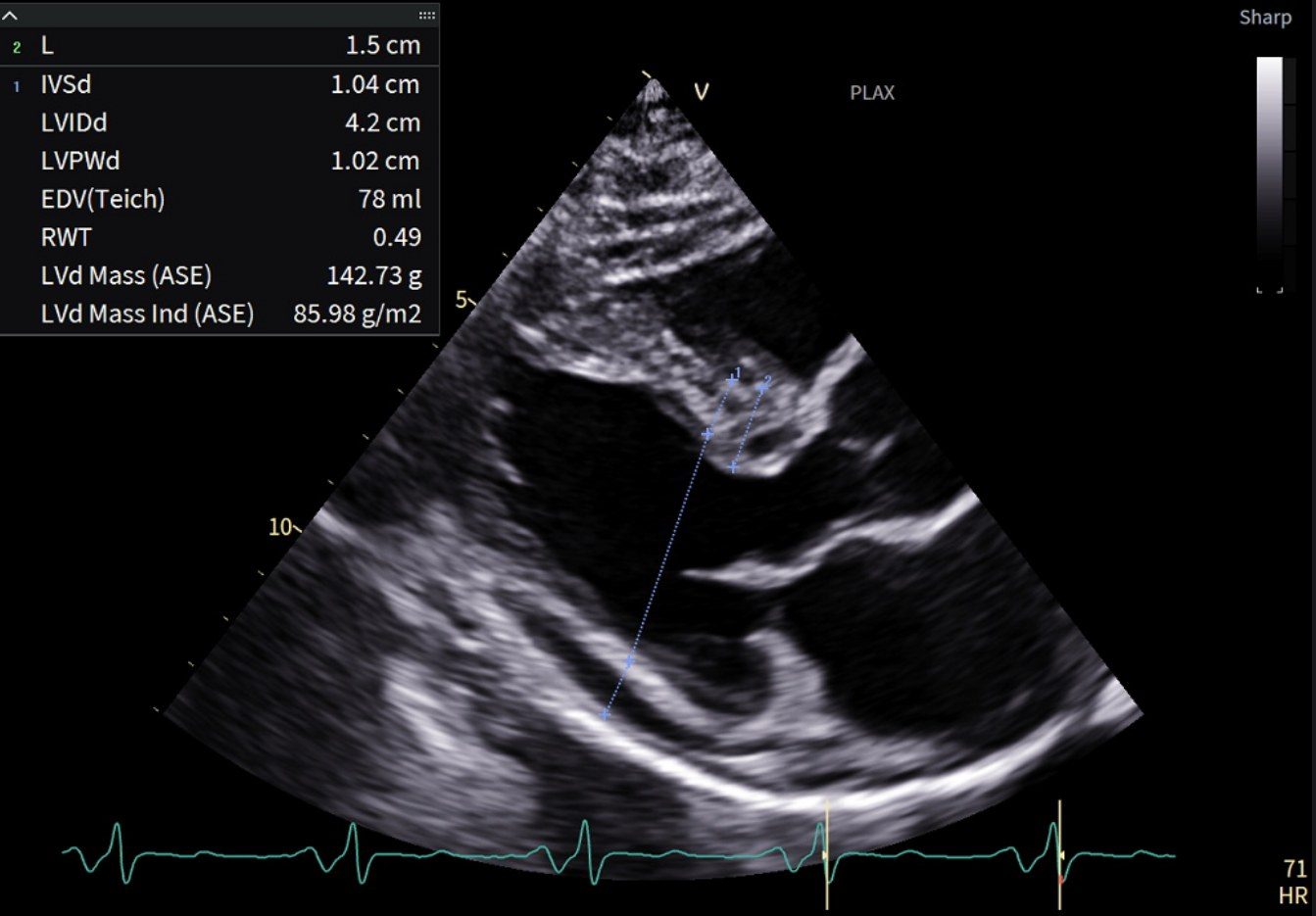
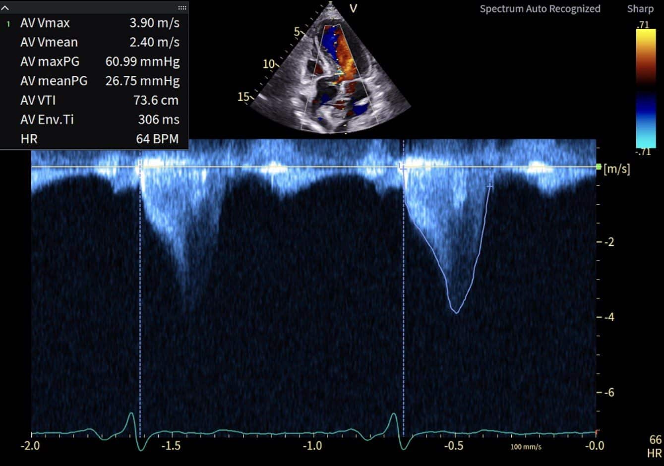
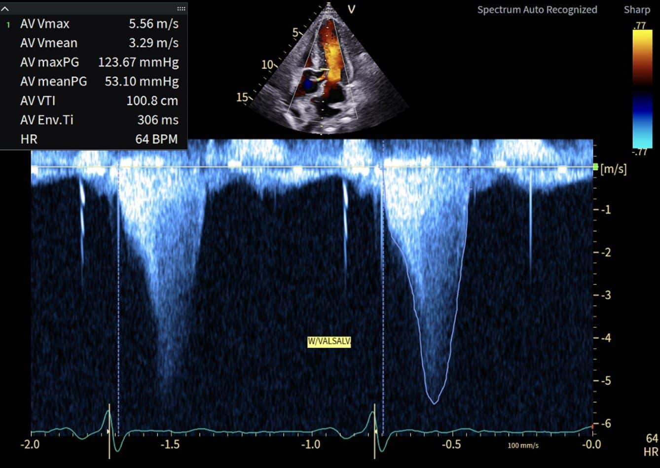
Post-treatment cine loops
She felt substantially better, although significant resting and provoked obstruction remained.
Case 2: a gradient above 200 mmHg with few reported symptoms
A woman in her late 60s was referred in 2025 after a loud systolic murmur was noted. She reported few cardiac symptoms during her predominantly sedentary daily routine. Her background included hypertension, type 2 diabetes and hypercholesterolaemia. CT coronary angiography showed a calcium score of 182, severe proximal RCA disease and a high-risk mid-LAD plaque.
During a low-intensity, manually adjusted treadmill protocol, she exercised for five minutes, achieving an estimated 3.75 METs and experiencing dyspnoea without chest pain. Her limited habitual activity may therefore have masked exertional symptoms.
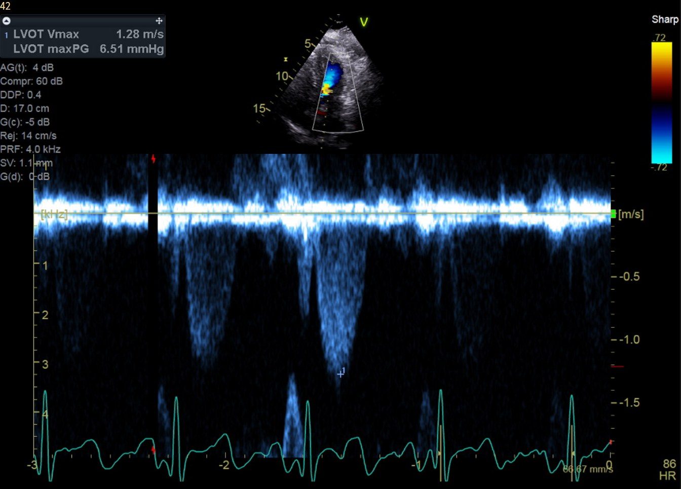
In 2022, resting LVOT velocity was approximately 1.28–1.29 m/s, with peak gradients of 6–7 mmHg.
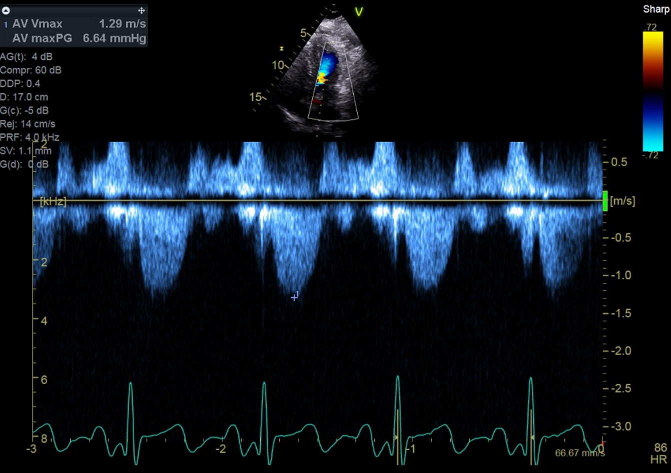
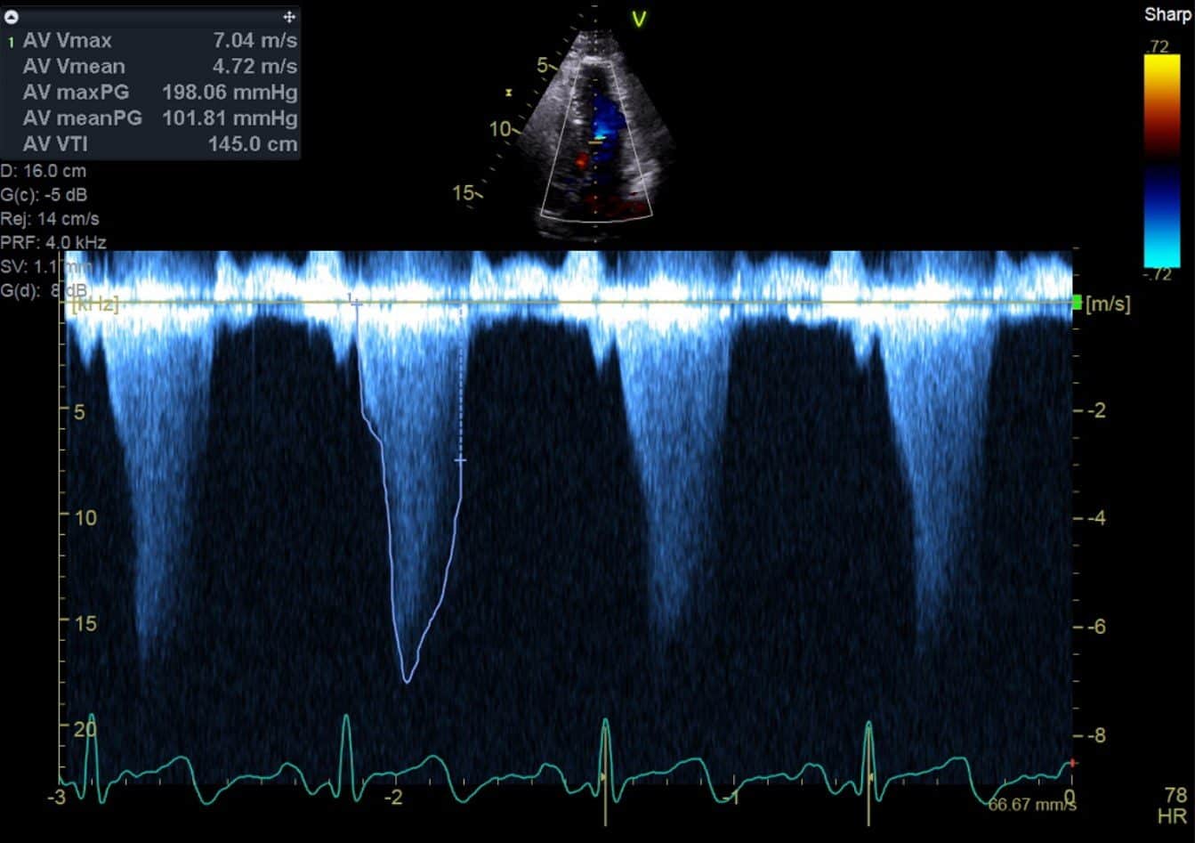
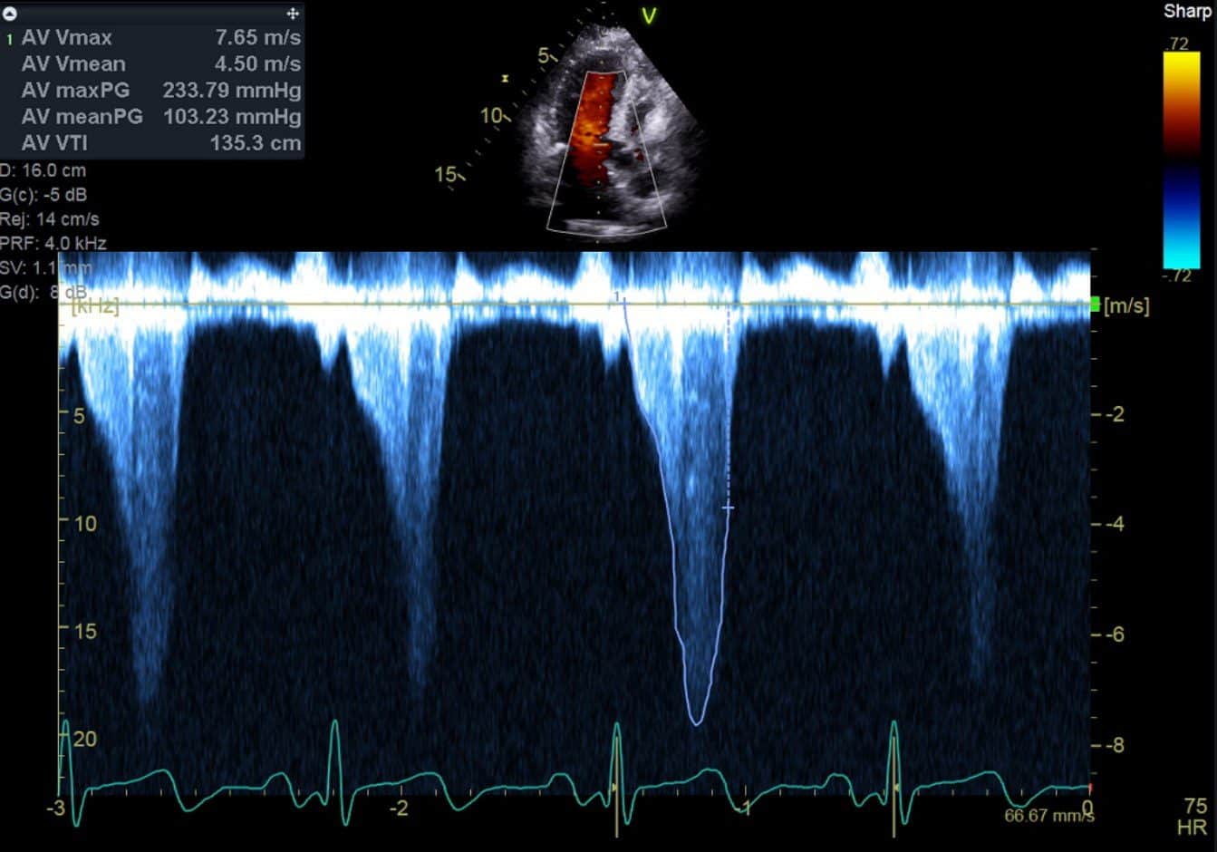
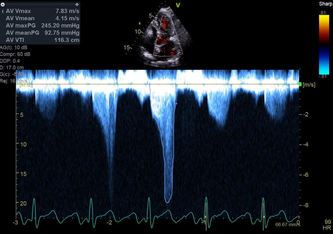
In 2025, resting CW recordings showed 7.04 m/s in the apical four-chamber view and 7.65 m/s in the three-chamber view, corresponding to peak gradients of 198 and 234 mmHg. The gradient reached 245 mmHg (7.83 m/s) after exercise and 256 mmHg (8.0 m/s) with Valsalva.
Her limited reported symptoms should be interpreted in the context of low habitual activity and dyspnoea during low-workload exercise. Her coexisting coronary disease also matters when interpreting exertional symptoms.
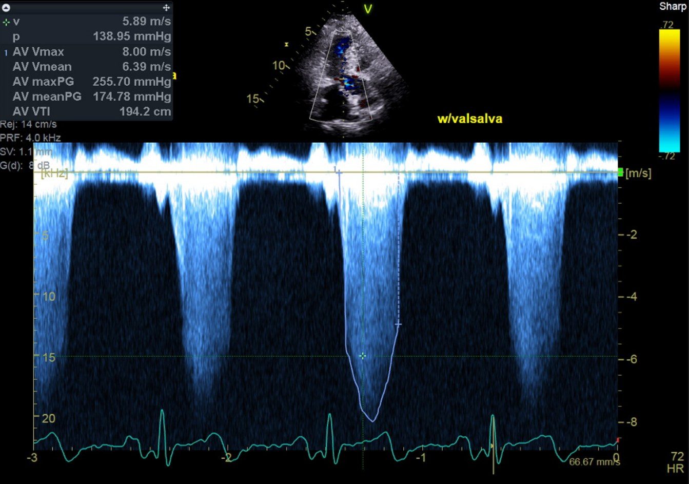
Why this signal is very unlikely to be mitral regurgitation
At velocities of 7–8 m/s, the first question is whether CW Doppler has captured the LVOT jet or mitral regurgitation (MR). In this patient, the findings strongly favour an LVOT origin.
Review of the full examination showed no appreciable MR, or at most a trivial jet. The accompanying colour loop shows no substantial regurgitant jet entering the left atrium. The mitral leaflets were otherwise normal apart from SAM.
The CW recordings show the late-peaking, dagger-shaped pattern of dynamic obstruction. As the LVOT narrows during systole, velocity rises towards a late peak. This shape supports the LVOT interpretation, although SAM-related MR can also begin late in systole.11
The Valsalva recording also contains a substantial, reproducible CW signal. Trivial MR usually produces a faint or incomplete envelope, so the density adds weight to the colour findings. Density depends on gain and beam alignment, however, and cannot identify the source on its own.13
Taken together, the absence of appreciable MR on the full examination, SAM and the late-peaking, dagger-shaped CW envelope strongly favour an LVOT origin and make MR contamination highly unlikely in this patient. Both MR and LVOT flow occur during systole. Although MR may begin before aortic ejection and persist after aortic valve closure, SAM-related MR can begin in mid-to-late systole, so timing and envelope duration are supporting features rather than definitive discriminators.11
The labels “AV Vmax” and “AV maxPG” on the screenshots reflect the machine’s measurement preset. These recordings were acquired through the dynamic LVOT jet; the labels do not indicate aortic stenosis.
Cardiac MRI findings
Cardiac MRI showed a maximum basal anteroseptal thickness of 15 mm with a non-sigmoid septal contour and a posterior wall of normal thickness, normal LV mass (55 g/m²), preserved biventricular function and normal atrial size. LGE occupied less than 5% of myocardial mass, at the RV insertion points and as a linear subepicardial band in the basal inferoseptum; no mid-wall LGE was reported in the thickened segment.
Native T1 was mildly elevated at approximately 1100 ms (local reference 960–1030 ms) and T2 was normal. Elevated native T1 indicates abnormal myocardial tissue composition but occurs in both HCM and hypertensive heart disease. The overall CMR pattern was not typical of cardiac amyloidosis.6,12
A negative genetic panel
Her cardiomyopathy panel found no medically significant variant. A negative result does not exclude HCM: sarcomere variants are identified in a minority of patients diagnosed at this age without a family history. Whether the septal thickening is better explained by hypertension is addressed below.1,2,3
For patients with a clinical diagnosis of HCM, first-degree relatives still need clinical screening when the gene panel is negative. There is simply no identified pathogenic variant to use for predictive genetic testing.1,2
Response to metoprolol
On metoprolol 75 mg in the morning and 50 mg in the evening, her resting heart rate was approximately 58 bpm. In 2026, resting LVOT velocity was 3.08 m/s and the peak gradient was approximately 38 mmHg. With Valsalva, the gradient remained approximately 133 mmHg (5.77 m/s). At follow-up, she reported few symptoms during her usual daily activities.
The large fall in the resting gradient is consistent with the dynamic nature of the obstruction. Serial comparisons should also account for heart rate, blood pressure, volume status and the way provocation was performed.
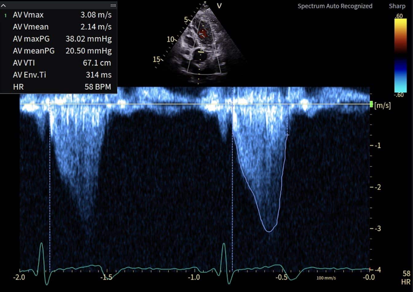
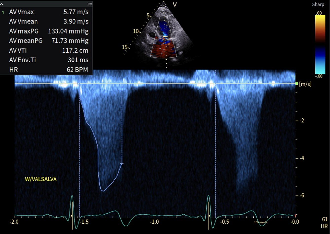
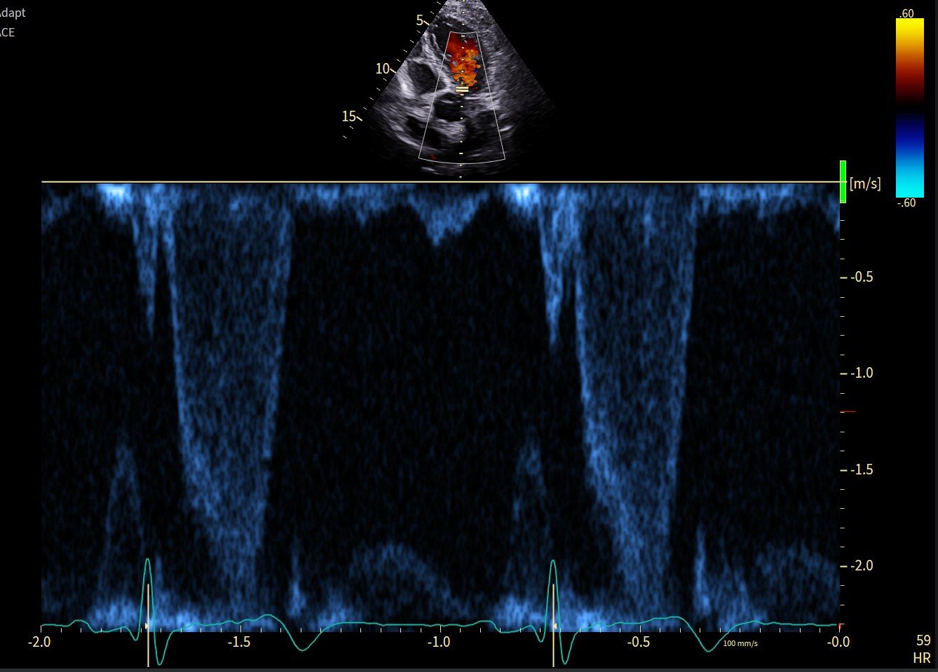
The resting gradient fell to 38 mmHg, but Valsalva still produced 133 mmHg. A lower resting gradient does not mean the obstruction has disappeared.
HCM or hypertensive basal septal remodelling?
In an older patient with hypertension, focal basal septal thickening may reflect HCM or age- and pressure-related remodelling. A thickness of 15 mm meets the numerical HCM threshold only when another cause does not adequately explain the hypertrophy. Severe LVOT obstruction alone does not settle the diagnosis.1,2
| Finding | This patient | Interpretation |
|---|---|---|
| Septal contour | Asymmetric septal thickening without a prominent sigmoid basal bulge | Less typical of an isolated age-related sigmoid septal bulge, but does not exclude hypertensive remodelling.16 |
| Posterior wall | Not thickened | Confirms an asymmetric pattern, which is common in HCM but also occurs in a minority of hypertensive patients.16 |
| Septal thickness | 15 mm basal anteroseptum | Meets the numerical HCM threshold; the diagnosis still requires that loading conditions do not adequately explain the hypertrophy.1,2 |
| Blood-pressure history | No history of severe or persistently uncontrolled hypertension | Weakens, but does not exclude, a hypertensive explanation for 15 mm of septal thickening. |
| LV mass | Normal: 55 g/m² | Normal mass is expected with focal hypertrophy and does not distinguish the two. |
| SAM and severe LVOTO | Present | Establishes obstructive physiology, but not its underlying cause. |
| Native T1 | 1100 ± 50 ms; reference 960–1030 ms | Mildly elevated, indicating abnormal tissue composition; seen in both HCM and hypertensive heart disease. |
| LGE | <5%; insertion points and linear subepicardial basal inferoseptum | Indicates a small burden of replacement fibrosis. Insertion-point LGE is non-specific, and no mid-wall LGE was reported in the thickened segment, which would have favoured HCM. |
| Genetic testing | No medically significant variant | Neither excludes HCM nor supports a hypertensive cause. Among patients with HCM, variant yield is low with later age at diagnosis, no family history and non-reverse-curvature septal morphology.3,15 |
Clinical interpretation (author): On balance, I favour HCM in this patient. She has no history of severe or persistently uncontrolled hypertension, and my review of the imaging shows asymmetric septal thickening without a prominent sigmoid basal bulge. Together with the CMR specialist’s interpretation, these findings support HCM as the working diagnosis. However, septal shape and a normal posterior wall do not reliably exclude hypertensive remodelling, and some contribution from hypertension remains possible.1,2,16
Initial measures to reduce dynamic obstruction overlap, but the underlying diagnosis affects subsequent management in three ways: whether first-degree relatives need clinical screening, whether HCM-specific sudden-death risk tools (validated in HCM, not in hypertensive remodelling) apply, and eligibility for mavacamten, which in Australia requires a diagnosis of obstructive HCM.1,2,14
The two cases at a glance
| Feature | Case 1 (late 50s) | Case 2 (late 60s) |
|---|---|---|
| Earlier resting gradients | 2023: LVOT 1.3–1.4 m/s; peak 7–8 mmHg | 2022: LVOT 1.28–1.29 m/s; peak 6–7 mmHg |
| 2025 presentation | Dyspnoea and presyncope | Incidental murmur; few daily symptoms; dyspnoea during exercise testing |
| Coronary status | CAC 0; no CAD on CTCA | CAC 182; severe proximal RCA disease, high-risk mid-LAD plaque |
| Maximum septal thickness | 15 mm basal anteroseptum | 15 mm basal anteroseptum |
| 2025 peak gradient | 94 mmHg rest; 133 mmHg Valsalva | 198–234 mmHg rest; 245 mmHg post-exercise; 256 mmHg Valsalva |
| Heart rate at acquisition | 2023 ≈90; 2025 ≈89 rest; 2026 ≈64–66 | 2022 ≈86; 2025 ≈75–78 rest, 99 post-exercise; 2026 ≈58–61 |
| MRI fibrosis | Moderate patchy mid-wall LGE (not quantified) | <5% LGE; insertion-point and subepicardial basal inferoseptal enhancement |
| Genetics | TNNT2 VUS; brother reportedly affected | No significant variant identified |
| Metoprolol | 100 mg morning + 75 mg evening | 75 mg morning + 50 mg evening |
| 2026 treated gradient | 61 mmHg rest; 124 mmHg Valsalva | 38 mmHg rest; 133 mmHg Valsalva |
| Clinical response | Marked symptomatic improvement | Resting gradient markedly reduced; few symptoms reported during usual activities |
Why severe obstruction can occur with a 15 mm septum
The severity of LVOT obstruction depends on how narrow the outflow tract becomes during systole, not on septal thickness alone. Systolic anterior motion (SAM) is driven mainly by flow drag: ejection flow pushes the anterior mitral leaflet towards the septum. If the leaflet contacts the septum, the remaining opening can become extremely narrow. The Venturi effect—the fall in local pressure as blood accelerates through the narrowed outflow tract—may contribute, but is not considered the main mechanism initiating SAM.
Septal shape, mitral leaflet length and papillary muscle position all influence how readily SAM develops. A basal septal bulge can redirect flow towards the leaflet, while a small, vigorously contracting LV brings the leaflet and septum closer together. The resulting gradient reflects both the degree of narrowing and the flow through the remaining opening. Severe obstruction can therefore occur even with modest septal thickening, explaining how a 15 mm septum can coexist with a gradient above 200 mmHg.
The gradient also changes with ventricular filling and contractility. During the strain phase of Valsalva, reduced venous return makes the LV smaller and can increase obstruction. Metoprolol reduces contractility and slows the heart rate, allowing more time for filling and often reducing the resting gradient.
Both cases demonstrate this: resting gradients fell substantially with metoprolol—from 94 to 61 mmHg in Case 1 and from 198–234 to 38 mmHg in Case 2—but marked obstruction remained during Valsalva, with gradients of 124 and 133 mmHg, respectively.
Measure maximum septal thickness, assess SAM and mitral valve anatomy, and record the LVOT gradient at rest and with provocation.
What cardiac MRI adds
CMR is particularly useful when echocardiography leaves uncertainty about the diagnosis, the distribution of hypertrophy or the mitral and papillary anatomy. It can identify apical disease, aneurysms and features suggesting an alternative diagnosis.1,2
Tissue characterisation adds information that the gradient cannot provide. Extensive LGE, often defined as at least 15% of LV mass, can influence sudden-death risk discussions and decisions about an implantable cardioverter-defibrillator (ICD). LGE should be measured and interpreted with the other risk markers.1,2,5
Here, both patients had a 15 mm septum, but their fibrosis findings differed: moderate patchy LGE in the younger patient and less than 5% LGE in the older patient. Similar wall thickness and obstruction do not imply the same myocardial disease burden.
What the genetic results mean for the family
Genetic results are most useful when their implications are clear. A pathogenic or likely pathogenic variant can guide testing in relatives. A VUS needs review and sometimes family studies; a negative panel leaves no variant available for predictive testing.1,2
Clinical screening remains important in families with HCM when testing finds a VUS or no causative variant. The younger patient’s family history strengthens the reason to assess relatives. Neither result, by itself, determines treatment or an individual patient’s sudden-death risk.1,2,3
Sudden-death risk is assessed separately from the gradient
A very high LVOT gradient is not, on its own, an indication for an ICD. Risk assessment also considers unexplained syncope, family history of HCM-related sudden death, non-sustained ventricular tachycardia, maximum wall thickness, LV systolic function, apical aneurysm and fibrosis. The gradient contributes to some risk models, but remains one part of the assessment.1,2
The younger patient reported presyncope rather than loss of consciousness, so this should not automatically be counted as unexplained syncope in sudden-death risk assessment. Although the description of “moderate fibrosis” is helpful, it does not establish whether the extent meets the definition of extensive LGE. Clarification of the fibrosis burden in the MRI report would help refine sudden-death risk assessment.
Treatment is directed at symptoms and functional limitation
Non-vasodilating beta-blockers, such as metoprolol, bisoprolol or atenolol, remain the initial treatment recommended by the 2024 AHA/ACC and 2023 ESC guidelines for symptoms attributable to LVOTO. Dose titration should follow symptoms, heart rate, blood pressure and tolerability. At higher doses, fatigue or reduced exercise capacity may reflect limited heart-rate response as well as the underlying disease.1,2
Mavacamten and aficamten are cardiac myosin inhibitors used to treat symptomatic obstructive HCM. Key criteria for initial PBS access to mavacamten include age ≥18 years, NYHA class II–III symptoms, a peak LVOT gradient ≥50 mmHg at rest or with provocation, and LVEF ≥55%. Maximum end-diastolic LV wall thickness must be ≥15 mm, or ≥13 mm with familial HCM. Eligibility also requires prior treatment with both a beta blocker and a non-dihydropyridine calcium channel blocker, unless the relevant drug class is contraindicated or not tolerated.1,2,7,14
Septal reduction is generally considered when symptoms attributable to obstruction persist and the resting or physiologically provoked gradient is at least 50 mmHg. With a septum of 15 mm, procedural planning needs particular attention to mitral and subvalvular anatomy. An experienced HCM team can weigh medication, surgery and alcohol septal ablation against the patient’s symptoms, anatomy and preferences.1,2
Review medications and volume status as well. Dehydration, excessive diuresis and vasodilators can worsen obstruction. This is especially relevant when treating coexisting hypertension, diabetes or coronary disease. Medication choices need to account for both conditions.1,2
The aim is better symptoms and function. A persistently high provoked gradient does not automatically require another drug or a procedure.
Medications that may worsen LVOT obstruction
| Medication | Examples | Main concern |
|---|---|---|
| Nitrates | GTN, isosorbide mononitrate | Reduced venous return; hypotension. |
| ACE inhibitors / ARBs | Perindopril, valsartan | Afterload reduction may worsen obstruction. |
| Dihydropyridine CCBs | Amlodipine, nifedipine | Vasodilation may worsen obstruction. |
| PDE-5 inhibitors | Sildenafil, tadalafil | Vasodilation and hypotension. |
| Excessive diuresis | Furosemide, indapamide | Volume depletion reduces LV filling. |
| Positive inotropes | Digoxin, dobutamine | Increased contractility intensifies obstruction. |
| Verapamil | In selected high-risk settings | Potentially harmful with hypotension, severe resting dyspnoea or resting gradients >100 mmHg. |
Note: Treatment is individualised. Low-dose diuretics may be used cautiously for congestion; verapamil remains an option in appropriately selected patients.
Aficamten: 2026 update
Aficamten is a cardiac myosin inhibitor that reduces excessive contractility. The 2025 MAPLE-HCM trial compared aficamten with metoprolol as monotherapy in 175 adults with symptomatic obstructive HCM. At 24 weeks, peak oxygen uptake increased by 1.1 mL/kg/min with aficamten and fell by 1.2 mL/kg/min with metoprolol: a between-group difference of 2.3 mL/kg/min (95% CI 1.5–3.1; P<0.001). Symptoms and haemodynamics also favoured aficamten.8
At ESC Munich in August 2026, a post-hoc MAPLE-HCM analysis examined whether previous beta-blocker treatment influenced the result. Aficamten improved peak oxygen uptake relative to metoprolol in both groups: by 1.9 mL/kg/min in patients previously taking beta-blockers and 3.1 mL/kg/min in those who were not. There was no statistically significant interaction with prior treatment (P=0.133). This supports the original finding, while remaining a subgroup analysis of the same trial.9
ACACIA-HCM, also presented in Munich, studied 517 patients with symptomatic non-obstructive HCM. At 36 weeks, aficamten improved the Kansas City Cardiomyopathy Questionnaire clinical summary score by 3.0 points and peak oxygen uptake by 0.67 mL/kg/min compared with placebo. The average benefits were modest, and systolic dysfunction was more frequent: LVEF fell below 50% in 10.5% versus 0.8%. Serious adverse events occurred in 20.2% versus 14.7%; a reduction in cardiovascular events was not demonstrated.10
For these two patients, the obstructive-HCM evidence is the relevant comparison. Neither patient received a myosin inhibitor. The younger patient improved on metoprolol. In the older patient, few symptoms reported during daily activities should be interpreted alongside the dyspnoea documented during low-workload exercise. Decisions about treatment escalation should reflect her current functional status and the likely contributors to any limitation.9,10
- Severe LVOT obstruction can occur with a 15 mm basal septum and normal overall LV mass.
- A low resting gradient does not exclude latent obstruction. Reassess with provocation when symptoms or findings change.
- Interpret very high CW velocities alongside colour Doppler, SAM, signal timing and envelope shape. In the older patient, the combined findings make MR contamination highly unlikely.
- CMR and genetic assessment answer questions that the gradient cannot. Quantify fibrosis and interpret a VUS or negative panel in the family’s clinical context.
- Follow symptoms, exercise capacity and resting and provoked gradients. Assess sudden-death risk separately; gradient magnitude alone does not determine an ICD or septal reduction decision.
References
- Ommen SR, Ho CY, Asif IM, et al. 2024 AHA/ACC/AMSSM/HRS/PACES/SCMR Guideline for the Management of Hypertrophic Cardiomyopathy. Circulation. 2024;149:e1239–e1311. doi:10.1161/CIR.0000000000001250
- Arbelo E, Protonotarios A, Gimeno JR, et al. 2023 ESC Guidelines for the management of cardiomyopathies. Eur Heart J. 2023;44:3503–3626. doi:10.1093/eurheartj/ehad194
- Ho CY, Day SM, Ashley EA, et al. Genotype and lifetime burden of disease in hypertrophic cardiomyopathy: insights from the Sarcomeric Human Cardiomyopathy Registry (SHaRe). Circulation. 2018;138:1387–1398.
- ClinVar. NM_001276345.2(TNNT2):c.341C>T (p.Ala114Val), Variation ID 177633: conflicting classifications of pathogenicity. National Library of Medicine. Source
- Chan RH, Maron BJ, Olivotto I, et al. Prognostic value of quantitative contrast-enhanced cardiovascular magnetic resonance for the evaluation of sudden death risk in hypertrophic cardiomyopathy. Circulation. 2014;130:484–495.
- Dorbala S, Ando Y, Bokhari S, et al. ASNC/AHA/ASE/EANM/HFSA/ISA/SCMR/SNMMI expert consensus recommendations for multimodality imaging in cardiac amyloidosis. J Nucl Cardiol. 2019;26:2065–2123, with 2021 addendum.
- Olivotto I, Oreziak A, Barriales-Villa R, et al. Mavacamten for treatment of symptomatic obstructive hypertrophic cardiomyopathy (EXPLORER-HCM): a randomised, double-blind, placebo-controlled, phase 3 trial. Lancet. 2020;396:759–769.
- Garcia-Pavia P, Maron MS, Masri A, et al. Aficamten or metoprolol monotherapy for obstructive hypertrophic cardiomyopathy (MAPLE-HCM). N Engl J Med. 2025;393(10):949–960. doi:10.1056/NEJMoa2504654
- Dominguez F, et al. Efficacy of aficamten vs metoprolol according to pretrial beta-blocker treatment in obstructive hypertrophic cardiomyopathy: a post-hoc analysis of MAPLE-HCM. JACC Heart Fail. 2026; published online 29 August 2026. Presented at ESC Congress 2026, Munich. Conference summary
- Masri A, Maron MS, Bhatia A, et al. Aficamten for symptomatic nonobstructive hypertrophic cardiomyopathy (ACACIA-HCM). N Engl J Med. 2026; published online 28 August 2026. doi:10.1056/NEJMoa2603021
- Nagueh SF, Phelan D, Abraham T, et al. Recommendations for multimodality cardiovascular imaging of patients with hypertrophic cardiomyopathy: an update from the American Society of Echocardiography. J Am Soc Echocardiogr. 2022;35:533–569. Source
- Messroghli DR, Moon JC, Ferreira VM, et al. Clinical recommendations for cardiovascular magnetic resonance mapping of T1, T2, T2* and extracellular volume. J Cardiovasc Magn Reson. 2017;19:75. doi:10.1186/s12968-017-0389-8
- Zoghbi WA, Adams D, Bonow RO, et al. Recommendations for noninvasive evaluation of native valvular regurgitation. J Am Soc Echocardiogr. 2017;30:303–371. Source
- Pharmaceutical Benefits Scheme. Mavacamten (Camzyos): symptomatic obstructive hypertrophic cardiomyopathy, authority required listing. Australian Government Department of Health. Accessed September 2026. Source
- Binder J, Ommen SR, Gersh BJ, et al. Echocardiography-guided genetic testing in hypertrophic cardiomyopathy: septal morphological features predict the presence of myofilament mutations. Mayo Clin Proc. 2006;81:459–467.
- Rodrigues JCL, Amadu AM, Dastidar AG, et al. Comprehensive characterisation of hypertensive heart disease left ventricular phenotypes. Heart. 2016;102:1671–1679. doi:10.1136/heartjnl-2016-309576
Echocardiography and cardiomyopathy assessment at Westmead
Resting and exercise echocardiography, provoked LVOT gradient assessment and cardiac MRI coordination for patients with suspected HCM or unexplained LV hypertrophy.
Refer a patient