Introduction
Table of Contents
stress echocardiogram—also called a stress echo or exercise stress echocardiography—combines a standard transthoracic echocardiogram with exercise to evaluate cardiac function under increased workload. The test uses ultrasound imaging to capture moving pictures of the heart at rest and immediately after physical exertion, enabling cardiologists to assess how effectively the heart muscle pumps blood under stress. This non‑invasive investigation is designed to detect reductions in blood flow caused by coronary artery narrowing and to evaluate structural or functional abnormalities. By comparing resting and post‑exercise images, clinicians can determine whether further diagnostic testing or adjustments to management are warranted.
Indications for stress echo (Why you might require stress echocardiogram)
Diagnosis of coronary artery disease and ischemia – The principal application of stress echocardiography is to determine whether the myocardium receives adequate blood supply during exertion. Coronary stenoses may produce symptoms only when heart rate and blood pressure increase, and stress imaging can reveal perfusion abnormalities that are not evident at rest.
Evaluation of unexplained exertional symptoms – Stress echocardiography provides direct observation of a patient’s cardiopulmonary response to exercise. It helps distinguish cardiac causes of chest discomfort, dyspnoea or fatigue from non‑cardiac causes, such as pulmonary disease or deconditioning.
Assessment of known cardiac conditions – Clinicians use stress echo to assess the severity of valvular lesions (e.g., aortic stenosis), cardiomyopathies, heart failure, congenital heart disease and pulmonary hypertension. The test demonstrates whether the heart’s contractile reserve is preserved or impaired.
Determination of safe exercise capacity – Following myocardial infarction, cardiac surgery or in chronic heart disease, stress echocardiography guides exercise prescription and cardiac rehabilitation by establishing a safe level of exertion.
Detection of exercise‑induced arrhythmias – Continuous electrocardiographic monitoring during stress echocardiography can reveal arrhythmias that manifest only under stress, aiding diagnosis and management.
Monitoring therapeutic interventions – Repeating stress echocardiography after medical therapy, angioplasty or coronary bypass surgery can demonstrate improvement in perfusion or ventricular function and inform further treatment.
Pre‑operative cardiac risk assessment – Prior to major non‑cardiac surgery, stress echocardiography may be used to evaluate myocardial reserve and stratify perioperative risk.
How to prepare for stress echocardiogram
To ensure a smooth stress echocardiogram experience, follow these preparation tips:
Clothing and footwear – Patients should wear comfortable, loose‑fitting clothing appropriate for exercise and supportive, non‑slip shoes. Trousers or shorts and a lightweight top allow the technologist to position electrodes and the ultrasound transducer effectively.
Fasting – A light meal may be taken earlier in the day, but it is generally advisable to avoid eating or drinking anything other than water for approximately three to four hours before the examination to minimise nausea or stomach discomfort during exertion.
Medication management – Routine medications should not be discontinued without medical advice. Patients should discuss all prescription and non‑prescription medicines with their physician in advance, as some agents—including beta‑blockers or caffeine‑containing remedies—may require adjustment.
Caffeine and nicotine – Caffeine (in coffee, tea, energy drinks and chocolate) and nicotine (from cigarettes, e‑cigarettes or patches) can alter heart rate and blood flow. These substances are usually avoided for at least 24 hours prior to a stress echocardiogram.
Alcohol and stimulants – Alcoholic beverages should be avoided for at least 24 hours before the test because they can affect the heart’s response to exercise. Patients should also abstain from other stimulants, including certain cold remedies or herbal supplements, unless approved by their doctor.
Removal of personal items – Jewellery, watches and electronic devices may interfere with the ultrasound equipment and should be removed before the examination. Bringing a list of current medications and allergies can assist the healthcare team in providing safe care.
The Stress Echocardiogram Procedure: Step-by-Step

The stress echocardiogram procedure consists of several stages. Here’s a step-by-step breakdown of the process:
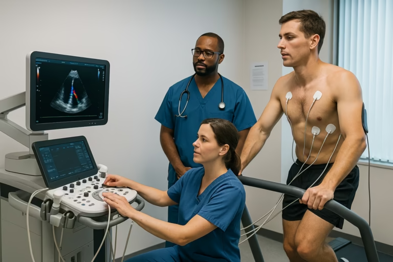
Baseline imaging – Before exercise begins, a cardiac sonographer acquires echocardiographic images of the heart at rest. A water‑based gel is applied to the chest to enhance ultrasound transmission, and a transducer is positioned to visualise the heart’s chambers, valves and overall pumping function.
Exercise phase – The patient then exercises on a treadmill or stationary bicycle to increase heart rate and cardiac workload. The intensity (speed and incline or resistance) is gradually increased following a standard protocol. Throughout this phase, the medical team continuously records blood pressure, heart rate and electrocardiographic data and notes any symptoms of discomfort.
Immediate post‑exercise imaging – While the heart rate remains elevated, the patient returns promptly to the examination table for repeat echocardiographic imaging. Capturing cardiac motion at peak stress allows direct comparison with the baseline images.
Recovery and monitoring – After the second set of images, the patient rests while staff monitor vital signs and symptoms until heart rate and blood pressure return to baseline.
Interpretation and reporting – A cardiologist reviews the ultrasound images and physiological data to identify any abnormalities in wall motion, valve function or blood flow. The findings guide decisions about additional testing, medical therapy, lifestyle modification or procedural intervention.
Typical images of a stress echocardiogram
Normal Stress Echocardiogram: In the following video, we can see a normal stress echocardiogram. The two images on the left show the heart at rest, with a heart rate of about 70 beats per minute. The two images on the right show the same images after exercise with a heart rate of about 120 beats per minute. We can see an excellent contraction of the heart muscle post-exercise with no lagging in any section.
Abnormal Stress Echocardiogram: In this video, again on the left side, we can see the heart at rest and on the right, the heart after exercise. However, in this instance, there is an area of the heart that doesn’t contract properly after exercise, where the arrows are pointing. This is due to a severe blockage in one of the main arteries of the heart, the LAD, which was confirmed with a coronary angiogram.
Understanding the Results: What to Expect
The possible outcomes of a stress echocardiogram can vary, and they may impact your treatment plan differently depending on the findings. At Heartcare Sydney, we discuss the results with the patient immediately after the test. Here are the possible results and their implications:
Normal response – A normal stress echocardiogram demonstrates uniform wall motion and ventricular contraction during peak stress without evidence of regional ischaemia, arrhythmia or valvular dysfunction. In such cases, additional investigation or modification of therapy is usually unnecessary.
Abnormal response – The study is considered abnormal when there is evidence of inducible ischaemia, exercise‑induced arrhythmias or abnormal valve function. Localised wall‑motion abnormalities appearing only under stress suggest significant coronary artery disease. Management decisions depend on the severity and distribution of these findings and may include pharmacological treatment, lifestyle modification, percutaneous coronary intervention or surgery.
Inconclusive study – Occasionally, the test does not yield definitive information because the patient fails to reach the target heart rate, image quality is suboptimal or the stress‑induced changes are subtle. In such circumstances, alternative diagnostic modalities—such as pharmacological nuclear stress testing, myocardial perfusion imaging or coronary computed tomography angiography—may be recommended to clarify the diagnosis.
Potential Risks and Complications of Stress Echocardiograms
While a stress echocardiogram is generally considered safe and non-invasive, there are some potential risks and complications associated with the test:
Arrhythmias – Transient tachycardia or irregular heart rhythms may develop during the exercise phase. These disturbances usually resolve when exertion ceases, although rare cases may require medical intervention.
Blood pressure changes – Exercise can cause blood pressure to rise or fall, leading to lightheadedness or dizziness. Such symptoms typically subside during the recovery period.
Chest pain or dyspnoea – Angina or shortness of breath may occur in individuals with underlying coronary artery disease or other cardiac conditions. The test is terminated promptly if significant symptoms develop, and appropriate treatment is initiated.
Myocardial infarction – An acute heart attack is an exceedingly uncommon but serious risk, particularly in patients with advanced coronary disease. Emergency protocols are in place to manage this event.
Overall, the incidence of serious complications is very low. Continuous monitoring allows the medical team to stop the procedure and provide care if concerning changes occur.
What are the alternative tests to Stress Echocardiogram?
There are alternative diagnostic methods available for patients who cannot undergo the standard treadmill stress echocardiogram due to mobility issues, health limitations, or other concerns. One such option is the pharmacological stress test, which utilizes medication to simulate the effects of exercise on the heart:
Pharmacological stress testing – For patients unable to perform adequate exercise because of musculoskeletal or respiratory limitations, pharmacological agents can simulate the cardiac workload. Vasodilators such as adenosine, dipyridamole or regadenoson dilate the coronary arteries and increase blood flow. Nuclear imaging techniques (SPECT or PET) are often used in conjunction to assess myocardial perfusion at rest and during stress. Continuous electrocardiographic and blood‑pressure monitoring is maintained throughout the procedure.
Dobutamine stress echocardiography – In patients who cannot exercise, intravenous administration of dobutamine—a beta‑adrenergic agonist that increases heart rate and contractility—provides an alternative. Echocardiographic images are obtained at rest and during graded doses of dobutamine to evaluate wall motion and ventricular function under stress. This method offers similar diagnostic information without requiring physical activity.
These alternative methods allow us to assess a patient’s heart function and blood flow in response to stress, even when traditional exercise-based stress testing is not feasible. It’s essential to discuss your specific situation to determine the most appropriate diagnostic method for your needs.
Can I have a CT instead of a stress echocardiogram?
A common question patients raise is whether a computed tomography coronary angiogram (CTCA) can replace a stress echocardiogram. These two investigations serve different, complementary purposes. CTCA is an anatomical study that produces detailed images of the large coronary arteries and detects atherosclerotic plaque or narrowing, but it does not assess how the heart responds to physiological stress or whether an identified narrowing actually limits blood flow.
Stress echocardiogram, on the other hand, is a functional test. By inducing exercise and monitoring cardiac performance under increased workload, it reproduces exertional symptoms such as chest pain or dyspnoea, measures exercise capacity and shows whether the myocardium maintains normal motion and perfusion during stress. This method can also reveal microvascular coronary disease—reduced blood flow in small vessels that are not visible on CT imaging.
Because these tests yield different insights, they are not interchangeable; instead, they can complement each other. A normal CTCA may confirm that the major coronary arteries are clear, while a stress echocardiogram can elucidate persistent exertional symptoms, assess functional capacity and stratify future cardiovascular risk. The choice of one or both modalities depends on the patient’s clinical presentation, risk factors and the judgement of the treating cardiologist.
Frequently Asked Questions about Stress Echocardiograms
Here are some common questions and concerns related to stress echocardiograms:
How long does a stress echocardiogram take? – The full procedure, including preparation, exercise, imaging and recovery, generally lasts between 45 and 60 minutes. The period of active exercise itself is comparatively short, usually six to twelve minutes, depending on the protocol and the individual’s level of fitness.
Will I need more tests after a stress echo? – The need for further investigation depends on the outcome. A study that is normal and consistent with the patient’s symptoms may not require additional testing. However, if the results are abnormal or inconclusive, the clinician may recommend further evaluation, such as coronary angiography, nuclear perfusion imaging or cardiac computed tomography.
How strenuous is the test? – The exercise component can be physically demanding, but the protocol is tailored to the patient’s age and physical capability. Throughout the test, vital signs and symptoms are closely monitored, and the examination is stopped if significant discomfort, chest pain or other concerning signs develop.
Is there radiation exposure with a stress echocardiogram? – No. Stress echocardiography uses ultrasound technology and does not expose the patient to ionising radiation, unlike nuclear stress tests or computed tomography scans (CT scan).
Conclusion
Stress echocardiography is an integral diagnostic tool in contemporary cardiology. By evaluating cardiac function and perfusion under controlled physiological stress, it assists in detecting coronary artery disease, valvular abnormalities, heart failure and exercise‑induced arrhythmias.
This non‑invasive ultrasound‑based procedure carries a low risk profile and provides real‑time information that guides therapeutic decisions. Early identification of abnormal responses to stress enables clinicians to recommend appropriate interventions—ranging from lifestyle modification and pharmacological therapy to revascularisation or surgical correction—thereby improving cardiovascular outcomes.
Individuals who experience chest discomfort, dyspnoea, unexplained fatigue or other symptoms suggestive of heart disease should seek a professional medical evaluation to determine whether stress echocardiography or another form of cardiac assessment is warranted.
If you are in Westmead or the Parramatta region and need a stress echo, visit our dedicated service page at stress-echocardiogram-westmead for booking information and local details. For patients elsewhere, talk to your doctor about where to have your test and whether a stress echocardiogram is right for you.