What Is an Echocardiogram?
An echocardiogram — also called an echo or heart ultrasound — is a painless, non-invasive test that uses sound waves to create moving images of the heart. It assesses the heart chambers, pumping function, valves and blood flow, without radiation or injections.
At Heartcare Sydney in Westmead, echocardiograms are performed on site by a cardiac sonographer and reported by consultant cardiologist Dr Reza Moazzeni. For detailed information about what an echo can diagnose, the different types of echocardiography and when the test is recommended, read our Echocardiogram Explained guide.
What to Expect
| Preparation | No fasting or medication changes are normally required. Wear comfortable clothing. |
|---|---|
| During the test | You lie on your left side while the sonographer obtains images from the chest. Painless — at most mild pressure from the probe. |
| Duration | Approximately 30 minutes. |
| Afterwards | Return to normal activities immediately. The report is sent to your referring doctor within 24–48 hours. |
NSW's First GE Vivid Pioneer
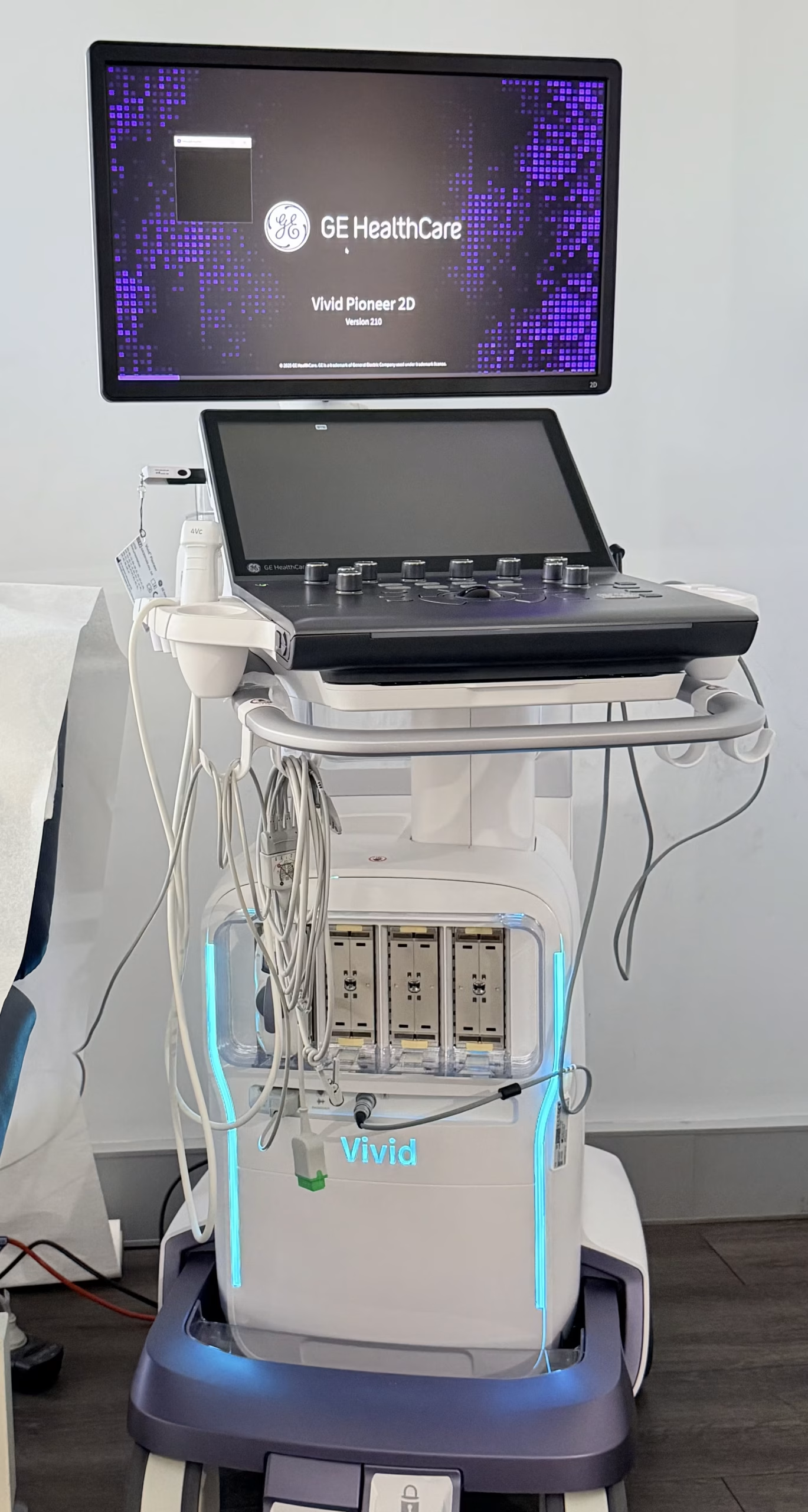
Heartcare Sydney uses NSW's first GE Vivid Pioneer cardiovascular ultrasound system. It provides advanced cardiac imaging with AI-assisted measurements, Doppler assessment and global longitudinal strain analysis when clinically appropriate — particularly valuable in technically challenging studies.
Fees, Billing and Referral Requirements
A stand-alone echocardiogram (no consultation) can be bulk billed for eligible patients — no out-of-pocket cost. Consultations are privately billed with a Medicare rebate. Stress echocardiography is not bulk billed. All fees are explained before your appointment.
A valid referral from your GP or specialist is required for all consultations and investigations, including stand-alone echocardiograms — this ensures you receive the appropriate Medicare rebate. Unsure about your referral? Call 02 8401 9598 and we will guide you.
Location, Transport and Parking
Fax: 02 8401 9599
9:00 am – 5:00 pm
Train & Light Rail
Parking
Referrals and Reporting
Send a Referral
Fax: 02 8401 9599
Please include current medications and the clinical indication
Reporting
All studies reported by Dr Reza Moazzeni, MD FRACP
Reports returned within 24–48 hours
Significant findings communicated directly to the referrer
Frequently Asked Questions
Do I need a referral for an echocardiogram in Westmead?
Yes — a referral from your GP or specialist is required for all consultations and investigations at Heartcare Sydney, including stand-alone echocardiograms. This ensures you receive the appropriate Medicare rebate. If you are unsure about your referral, call us on 02 8401 9598.
Can the echocardiogram be bulk billed?
If you attend for a stand-alone echocardiogram without a consultation, the test can be bulk billed for eligible patients — no out-of-pocket cost. Consultations are privately billed with a Medicare rebate, and stress echocardiography is not bulk billed. All fees are explained before your appointment.
Can I have an echocardiogram without seeing the cardiologist?
Yes. With a referral for an echocardiogram, you can attend for the test only. The study is performed by our cardiac sonographer, reported by Dr Moazzeni, and the report is sent to your referring doctor within 24–48 hours. A consultation can be arranged separately if your doctor recommends one.
How long does the test take, and is preparation required?
A standard resting echocardiogram takes approximately 30 minutes. No fasting or medication changes are normally required — simply wear comfortable clothing. If combined with a consultation, allow around 60 minutes.
Is an echocardiogram painful?
No — it is non-invasive and painless. It uses ultrasound waves, similar to a pregnancy scan, with no radiation or needles. At most you may feel mild pressure from the probe on your chest.
When will my doctor receive the result?
A detailed report is sent to your referring doctor within 24–48 hours. If your echocardiogram is combined with a consultation, Dr Moazzeni will usually discuss the findings with you on the same day.
Where is the clinic and where can I park?
We are at Office 33 (Level 3), 163–171 Hawkesbury Road, Westmead — opposite Westmead Hospital and about 200 metres from Westmead Station. There is no dedicated patient parking, but 2-hour street parking is available around Westmead Hospital. Public transport is recommended where possible.
What is the difference between a resting echo and a stress echo?
A resting echocardiogram images the heart at rest, assessing structure, valve function and pumping strength. A stress echocardiogram adds treadmill exercise to assess how the heart performs under stress, primarily to detect coronary artery disease. Both are available at our Westmead clinic — see our stress echocardiogram page for details.
Cardiologist-Reported Echo in Westmead
Opposite Westmead Hospital, 200 metres from Westmead Station. Bulk billed for eligible patients with a valid referral.
Book an Echocardiogram →or call 02 8401 9598