Unilateral leg swelling, where one leg increases in size relative to the other, often poses significant diagnostic challenges. It is less common and more difficult to diagnose than bilateral leg swelling, which is usually linked to heart failure and chronic venous insufficiency. The common causes of bilateral swelling cannot be assumed in a patient presenting with one swollen leg, and a deeper, more systematic investigation is required.
Categorising the swelling as acute or chronic is the essential first step. The pathologies responsible for unilateral leg swelling include venous stasis (deep vein thrombosis), malignancy, lymphoedema, trauma, and infection (such as cellulitis).
Acute versus chronic unilateral leg swelling
Acute causes
Typically associated with sudden onset and rapid progression. Infectious conditions such as cellulitis, and vascular issues such as deep vein thrombosis (DVT), are among the most common acute causes. Less common acute causes include malignancy and mass effect from compression of major veins (such as May-Thurner syndrome).
Chronic causes
Characterised by gradual onset and persistent symptoms. Post-thrombotic syndrome (also known as venous stasis syndrome) and secondary lymphoedema resulting from trauma or cancer treatment are common attributions for chronic unilateral leg swelling.
This post focuses on the less-well-understood chronic causes and presents two illustrative case studies that highlight the importance of comprehensive history-taking and physical examination. For the patient-facing version of this topic, see the pillar guide on leg swelling (lower limb oedema).


Acute deep vein thrombosis — the most important acute cause to exclude in any new unilateral leg swelling.
Case 1: Post-thrombotic syndrome
Current presentation
A 78-year-old man presents with persistent symptoms in his left leg — chronic skin discolouration, thickening, mild swelling, and a dull ache troubling him for over a decade. Examination reveals hyperpigmentation, lipodermatosclerosis, and patches of eczema — signs indicating a chronic venous origin.
Past medical history
A detailed history revealed an episode of acute deep vein thrombosis in the same leg twelve years earlier. The images below confirm a severe episode of DVT that required extensive hospitalisation and anticoagulation. His records note the development of venous ulcers, since healed, indicating a long-standing course of venous insufficiency. This presentation exemplifies a clinically significant form of post-thrombotic syndrome — a consequence of his earlier DVT.


Case 2: Traumatic lymphoedema
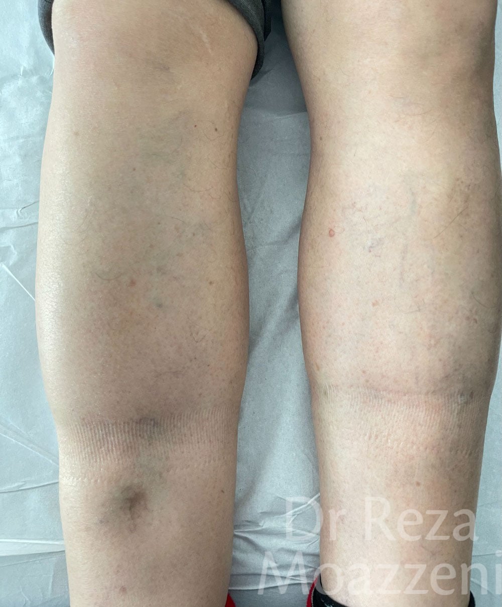
A 75-year-old man, recently diagnosed with a neuroendocrine tumour requiring chemotherapy and surgical intervention, was referred for evaluation of his swollen right leg. Despite presenting with pitting oedema — a sign often raising concern for heart failure — there were no accompanying features such as orthopnoea, paroxysmal nocturnal dyspnoea, or a decrease in exercise tolerance, which he maintained consistently over the years.
Further investigation, including an echocardiogram, showed no signs of heart failure (which is in any case uncommonly associated with unilateral swelling). The absence of lymphadenopathy or mass effect also ruled out other typical causes of lymphoedema.
Deeper history revealed that the swelling began following a severe car accident at age 30, which resulted in multiple fractures in the right leg and prolonged immobilisation. This long-standing history of trauma led to chronic unilateral leg swelling, identified as traumatic lymphoedema — a diagnosis confirmed through comprehensive history-taking and the exclusion of other conditions.
The differential: a clinician's reference
A structured differential, divided by time course. The "pitfall" column flags the errors most likely to delay diagnosis.
| Cause | Onset | Key features | Investigation | Pitfall |
|---|---|---|---|---|
| Acute | ||||
| Deep vein thrombosis | Hours to days | Calf pain, warmth, tenderness; risk factors present | Wells score, D-dimer, compression duplex US | A negative D-dimer in high pre-test probability does not exclude DVT |
| Cellulitis | Hours to a day | Red, warm, tender; often febrile; border may be indistinct | Clinical; bloods if systemic | Bilateral "cellulitis" is rarely infective — reconsider |
| Ruptured Baker's cyst | Sudden | Pain behind knee, bruising tracking down calf | Ultrasound | Mimics DVT — exclude DVT first |
| Acute arterial occlusion | Sudden | Pain, pallor, pulselessness, cold limb | Urgent arterial imaging | Limb-threatening — do not mistake for venous swelling |
| Compartment syndrome | Acute | Severe pain out of proportion, tense compartment | Compartment pressures; clinical | Surgical emergency |
| Gout / pseudogout | Hours | Acute monoarthritis, erythema; may mimic cellulitis | Joint aspiration, urate | Periarticular swelling misread as diffuse limb swelling |
| Chronic | ||||
| Post-thrombotic syndrome | Months to years post-DVT | Hyperpigmentation, lipodermatosclerosis, ulceration; prior DVT | Venous duplex US; history | Forgetting to ask about remote DVT |
| Chronic venous insufficiency | Years | Varicose veins, end-of-day heaviness; usually bilateral but can be asymmetric | Venous duplex US | Assuming bilateral — can be markedly one-sided |
| Lymphoedema (non-traumatic) | Gradual | Positive Stemmer sign, dorsum of foot involved; often post-cancer | Clinical; lymphoscintigraphy if uncertain | Excluding it because it pits early |
| Traumatic lymphoedema | Delayed, post-injury | History of significant limb trauma or surgery, sometimes decades earlier | Clinical; exclusion | Missing the remote trauma history |
| Neoplastic / mass effect | Subacute to chronic | Pelvic or proximal mass compressing venous/lymphatic return | Cross-sectional imaging | Overlooking malignancy in unexplained unilateral swelling |
| Chronic compartment syndrome | Exertional, chronic | Exercise-induced pain and tightness, resolves with rest | Dynamic compartment pressures | Mislabelled as musculoskeletal strain |
Discussion
Distinguishing acute from chronic unilateral leg swelling is essential for accurate diagnosis. Acute swelling may arise from infection (cellulitis — rapid onset of redness, warmth and tenderness) or vascular causes such as DVT, where thrombus obstructs venous return, causing swelling, pain and discolouration. Acute arterial occlusion is a critical cause demanding immediate attention because of the sudden loss of circulation and risk to the limb.
Chronic conditions such as post-thrombotic syndrome can develop after DVT, with symptoms typically emerging within six months but potentially appearing up to two years after the event. PTS affects 20–50% of patients following a DVT and is marked by pain, persistent oedema, skin discolouration and ulceration, reflecting the long-term consequences of venous damage.
Lymphoedema, often resulting from damage to the lymphatic system through surgery, radiation or trauma, leads to persistent swelling requiring meticulous management to prevent progression and complications. Traumatic lymphoedema — a specific subset — can occur after direct injury to lymphatic vessels or nodes, as in severe limb trauma, crush injuries, or penetrating wounds. The resulting lymphatic dysfunction causes persistent swelling, fibrosis, and an increased risk of infection. Diagnosis requires a high index of suspicion and a detailed history, as the onset of symptoms may be substantially delayed following the initial injury.
Conclusion
With its diverse aetiologies, unilateral leg swelling poses a significant diagnostic challenge. A systematic approach — thorough history and physical examination, divided by acute versus chronic onset — is critical in distinguishing the causes. Recognising the unique features of conditions such as post-thrombotic syndrome and traumatic lymphoedema is vital for accurate diagnosis and targeted management. Comprehensive history-taking, as both cases illustrate, frequently provides the decisive clue.